
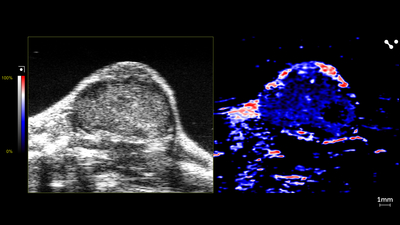
Oyx-hemo Imaging of Mouse Tumor
Oxy-hemo image of a mouse tumor showing shear wave elastography, scanned using a UHF29x transducer.

Contrast Imaging of Mouse Tumor
Contrast image of a mouse tumor showing shear wave elastography, scanned using a UHF29x transducer.
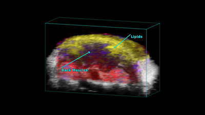
Mouse Scapular
PA-Mode 3D unmixing of a mouse scapular, highlighting the back muscles and lipids. Captured using a UHF29x transducer.

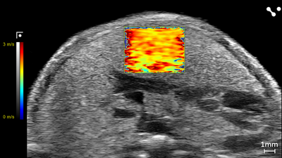
Shear Wave Elastography: NAFLD/MASLD Mouse - Velocity
Liver in a NAFLD/MASLD Mouse showing Velocity (m/s) through Shear Wave Elastography scanned using a UHF29x transducers on a Vevo F2 system.
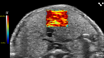
Shear Wave Elastography: NAFLD/MASLD Mouse - Stiffness
Liver in a NAFLD/MASLD Mouse showing Stiffness (kPa) through Shear Wave Elastography scanned using a UHF29x transducers on a Vevo F2 system.
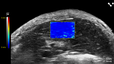
Shear Wave Elastography: Control Mouse - Velocity
Liver in a Control Mouse showing Velocity (m/s) through Shear Wave Elastography scanned using a UHF29x transducers on a Vevo F2 system.
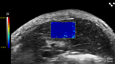
Shear Wave Elastography: Control Mouse - Stiffness
Liver in a Control Mouse showing Stiffness (kPa) through Shear Wave Elastography scanned using a UHF29x transducers on a Vevo F2 system.
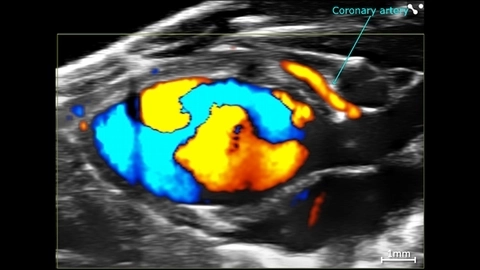
Cardiac Flow Dynamics and Coronary Artery
Cardiac flow dynamics and Coronary Artery visualized with Color Doppler EKV on the Vevo F2 System.
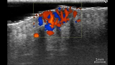
Color Doppler in Basal Cell Carcinoma Lesion
Color Doppler showing blood flow within a basal cell carcinoma lesion in a patient using ultra high frequency ultrasound on the Vevo MD.
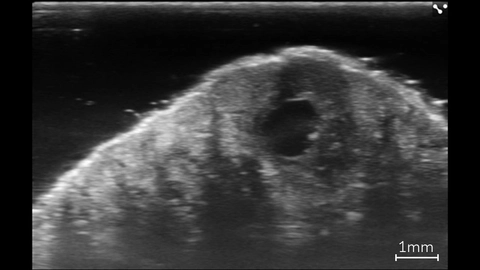
Basal Cell Carcinoma Lesion
High resolution image of a basal cell carcinoma lesion in a patient, imaged with the Vevo MD.
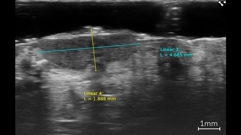
Linear Measurements of a Basal Cell Carcinoma
Linear measurements of a basal cell carcinoma in a patient using ultra high frequency ultrasound on the Vevo MD.

Canine eye in vivo
Entire canine eye visualized in vivo with the Vevo F2, scanned using a UHF22x transducer.
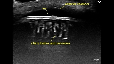
Canine eye angle of the anterior chamber
Canine eye imaged in vivo on the Vevo F2 scanned using the UHF71x.
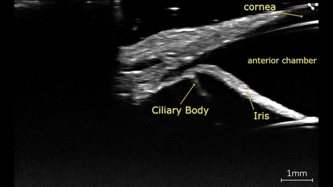
Canine eye, Ciliary body and processes
In vivo canine eye with ciliary processes, imaged with high-frequency on the Vevo F2 scanned using the UHF71x.
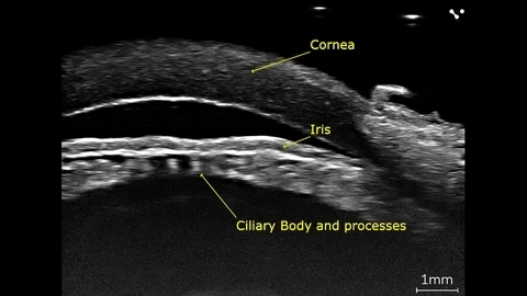
Porcine eye, Ciliary body and processes
Porcine eye imaged ex-vivo using the Vevo F2 with a UHF71x transducer.
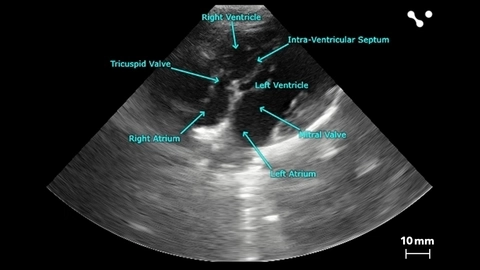
Apical 4 Chamber View of a Beagle
Apical 4 chamber view of a beagle scanned using a P5-1 transducers on the Vevo F2. Images courtesy of Drs. Kenneth Hoyt and Jay Griffin at Texas A&M University.
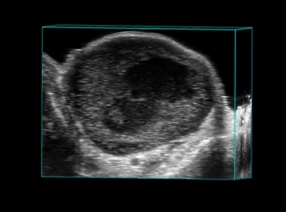
Breast tumor in 3D
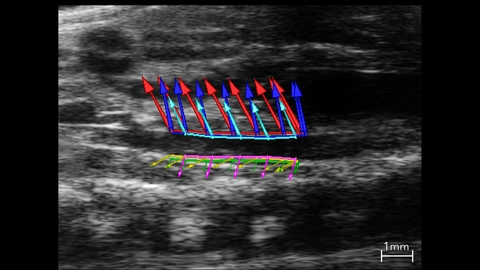
Abdominal Aorta Used In Strain Analysis
B-mode image of the abdominal aorta with vessel wall velocity vector arrows used in strain analysis.
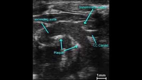
Atherosclerotic Plaque in an Aortic Arch
2D image showing atherosclerotic plaques in a mouse aortic arch.