
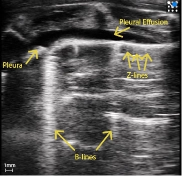
Rat Pleural Effusion
Rat with pleural effusion and pleural thickening. Clear B-Lines can be seen as well as Z-Lines.
Image courtesy of : M.Sc. Niklas Hegemann, Kübler lab, Institute of Physiology, Charité-Universitätsmedizin Berlin & Dr. Jana Grune, Nahrendorf lab, Center for Systems Biology, Massachusetts General Hospital.
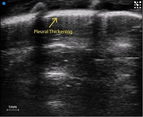
Pleural thickening
Substantial pleural thickening in the rat lung.
Image courtesy of : M.Sc. Niklas Hegemann, Kübler lab, Institute of Physiology, Charité-Universitätsmedizin Berlin & Dr. Jana Grune, Nahrendorf lab, Center for Systems Biology, Massachusetts General Hospital.
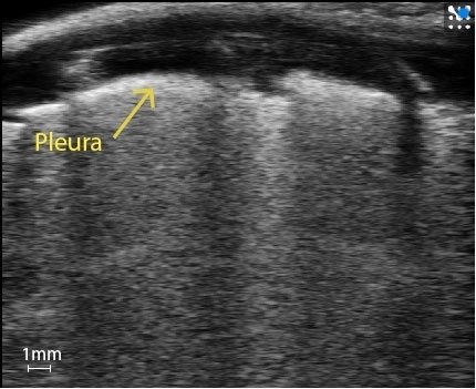
Murine White Lung
Mouse lung covered in B-Lines, creating white lung.
Image courtesy of : M.Sc. Niklas Hegemann, Kübler lab, Institute of Physiology, Charité-Universitätsmedizin Berlin & Dr. Jana Grune, Nahrendorf lab, Center for Systems Biology, Massachusetts General Hospital.
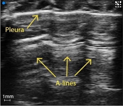
Healthy mouse lung
Healthy naive mouse lung with echogenic pleura and A-Lines ringing down.
Image courtesy of : M.Sc. Niklas Hegemann, Kübler lab, Institute of Physiology, Charité-Universitätsmedizin Berlin & Dr. Jana Grune, Nahrendorf lab, Center for Systems Biology, Massachusetts General Hospital.
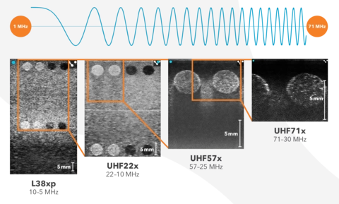
Vevo F2 Range of Frequencies
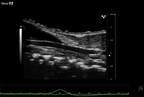
Mouse carotid artery
Scanned using a UHF71x transducer on the Vevo F2.
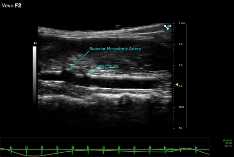
Mouse abdominal aorta
Scanned using a UHF46x transducer on the Vevo F2.

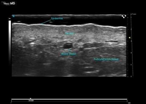
High Resolution Ultrasound Dermal Imaging
Scanned using a UHF70 transducer on the Vevo MD.
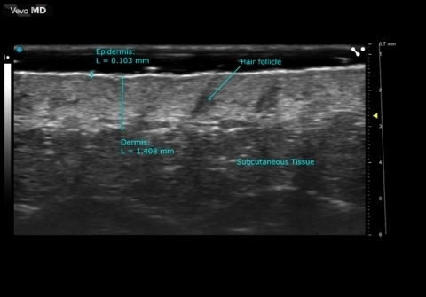
Facial Skin
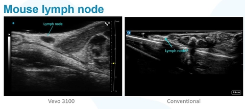
Mouse lymph node - Vevo 3100 vs. Conventional
The difference is obvious. Lymph node on the left was acquired using ultra high frequency ultrasound (Vevo 3100) whereas the image on the right is the same lymph node using conventional ultrasound.
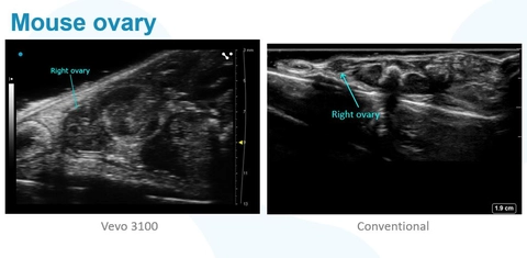
Mouse Ovary - Vevo 3100 vs. Conventional
Mouse ovary acquired using ultra high frequency ultrasound (Vevo 3100) against an image of the same mouse ovary using a conventional ultrasound system.
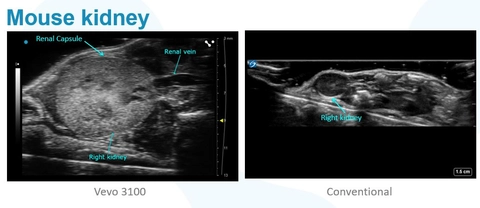
Mouse Kidney - Vevo 3100 vs Conventional
What you see using the Vevo 3100 compared with what you can see using a conventional ultrasound system is shown here. See the mouse kidney in great detail using ultra high frequency ultrasound imaging.
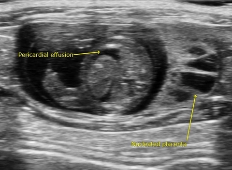
Pericardial Effusion - Spina Bifida Rat Model
Spina bifida is a neural tube defect that results in incomplete closing of the spinal cord. Spina bifida is associated with abnormalities in the cerebellum and cisterna magna during fetal development. This image acquired using the Vevo 3100.