Cardiovascular Biology
Preclinical Echocardiography for Cardiology Research
Ultra-high frequency (UHF) ultrasound provides superior resolution and will have an incredible impact on cardiovascular research in your animal models.
Perform rapid, repeatable echocardiography in small to large animals in vivo with resolution down to 30 μm.
Vevo ultrasound imaging platforms provide the best temporal and spatial resolution compared to MRI, conventional ultrasound and CT. The Vevo F2xc system can perform preclinical echocardiography in animal models all the way from zebra fish and mice to pigs. Vevo technology has been established as a gold standard technology for mouse cardiac imaging, having been utilized in over 3,000 peer-reviewed publications.
With the Vevo imaging systems you can:
- Perform cardiovascular phenotyping for cardiac structure and function, ideal for stress echo and cardiotoxicity studies
- Image the heart in 4D (with Color or Power Doppler integration)
- Quantify diastolic dysfunction
- Evaluate early signs of cardiac dysfunction with cardiac strain (global or regional synchrony and deformation)
- Perform image-guided cardiac injections
- Monitor full animal physiology while imaging, including body temperature, ECG and respiratory rate
- Assess myocardial oxygen saturation
Request a Quote, Demo or Spec Sheet
Vevo Software
Vevo Strain 2.0
Early Detection of Myocardial Deformation using Speckle-tracking Software.
Strain is a more direct measurement of intrinsic myocardial contractility than global values such as ejection fraction or fractional shortening. However, the real power of cardiac strain comes in its ability to detect dysfunction earlier than standard functional parameters!
On-Demand Workshop
Virtual Advanced Cardio Symposium
Innovative science, advanced imaging techniques and impactful insights are shaping the future of cardiovascular research. If you missed this workshop, now's your chance to catch up.
Gallery

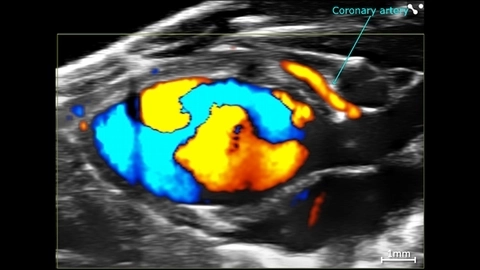
Cardiac Flow Dynamics and Coronary Artery
Cardiac flow dynamics and Coronary Artery visualized with Color Doppler EKV on the Vevo F2 System.
Apical 4 Chamber View of a Beagle
Apical 4 chamber view of a beagle scanned using a P5-1 transducers on the Vevo F2. Images courtesy of Drs. Kenneth Hoyt and Jay Griffin at Texas A&M University.


Color Doppler of Mouse Aortic Arch
Color Doppler image of an aortic arch in a female mouse, acquired using a UHF57x transducer on the Vevo F2.
Publications
TOP PAPER
Left atrial reservoir strain as a predictor of cardiac dysfunction in a murine model of pressure overload
Acta Physiologica
,
TOP PAPER
Endothelial cells drive organ fibrosis in mice by inducing expression of the transcription factor SOX9
Science Translational Medicine
,
TOP PAPER
In-ovo echocardiography for application in cardiovascular research
Basic Research in Cardiology
,
TOP PAPER
Echocardiography protocol: A tool for infrequently used parameters in mice
Frontiers in Cardiovascular Medicine
,
TOP PAPER
Comparative assessment of coronary physiology using transthoracic pulsed-wave Doppler and myocardial contrast echocardiography in rats
European radiology experimental
,
TOP PAPER
Embryonic echocardiography for assessment of congenital and functional cardiac defects
STAR Protocols
,
TOP PAPER
P2Y12 Inhibition in Murine Myocarditis Results in Reduced Platelet Infiltration and Preserved Ejection Fraction
Cells
,
TOP PAPER
Role of plakophilin-2 expression on exercise-related progression of arrhythmogenic right ventricular cardiomyopathy: a translational study
European Heart Journal
, Request a Quote or Demo