Splenic Vasculature in a Mouse
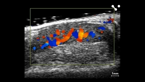
Color Doppler of Rat Kidney Vasculature
Color Doppler of rat kidney vasculature scanned using a UHF29x transducer.
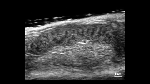
Rabbit Kidney
Rabbit kidney scanned using a UHF29x transducer.
Murine Spleen
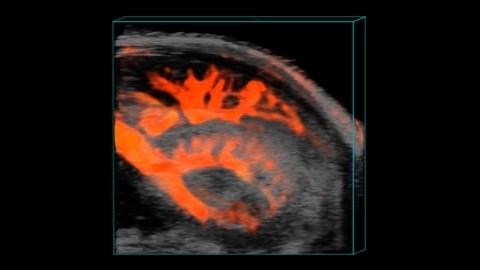
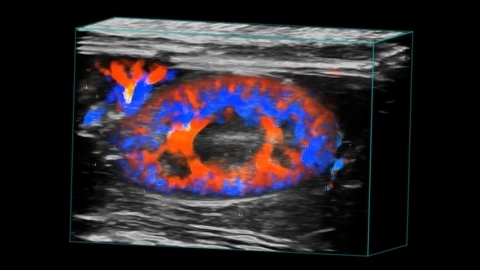
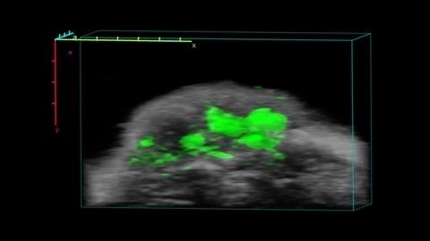
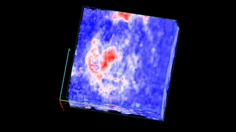
Murine Renal Vasculature in 3D
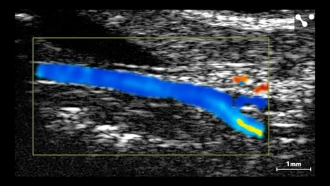
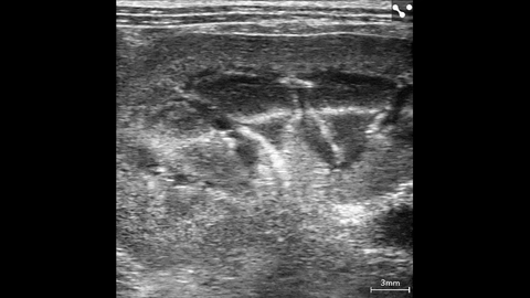
Murine Carotid Artery
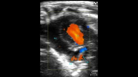
Murine Apical Color Doppler
Murine apical color Doppler scanned using a UHF46x transducer.
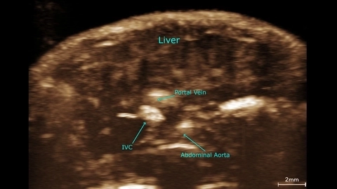
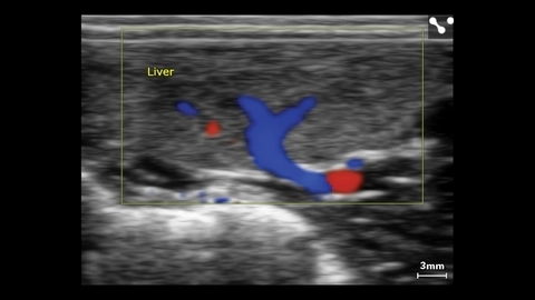
Mouse Liver with Major Vessels
Vascular perfusion in the mouse liver
Mouse liver contrast MIP scanned using a UHF29x transducer.
Mouse Kidney, Spleen and Pancreas
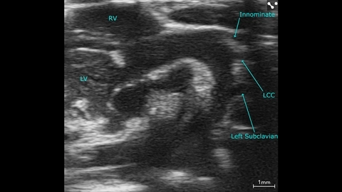
Mouse Aortic Arch
Mouse aortic arch scanned using a UHF57x transducer.
Parasternal Long Axis of Mouse Heart in 4D
Parasternal long axis of a mouse heart in 4D scanned using the UHF46x transducer.

Mitral flow in a small dog
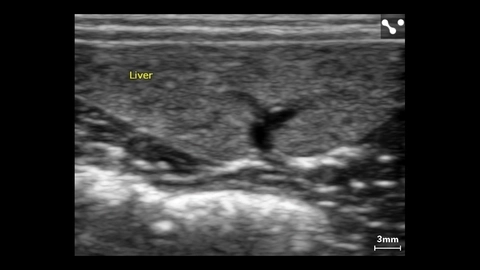
Liver Vasculature in a Mouse
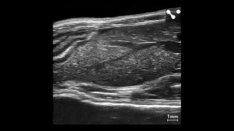
ICG localization in tumor
Cerebral Oxygenation after Carotid Artery Occlusion

Anterior Myocardium Oxygenation, High O2 stats in Red, Low in Blue
Liver Color Doppler in Small Dog
Liver Color Doppler of small dog scanned using a L38xp transducer.
Liver of Small Dog
Liver of small dog measured using a L38xp transducer.
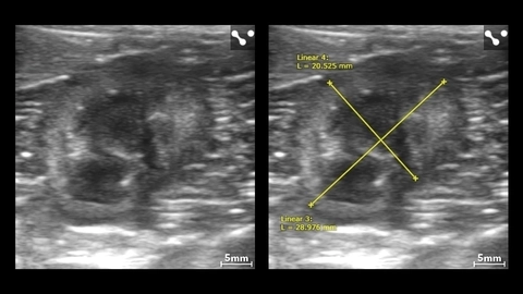
Left Kidney in Small Dog with Measurements
Left kidney in small dog with measurements imaged using the L38xp transducer.
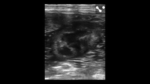
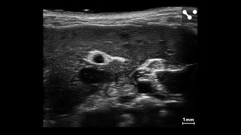
Kidney in a Small Canine