Developmental
Image mouse and rat embryos with superior resolution
The size, structural details and function of the placenta and developing embryo can be measured over multiple gestational time-points without having to sacrifice your animal.
With the Vevo imaging systems you can:
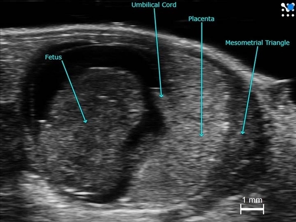
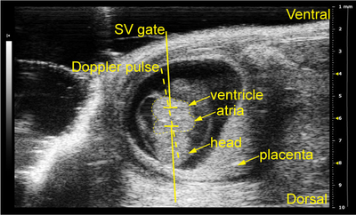
Mouse implantation site at mid-gestation.
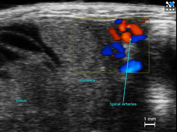
Spiral arteries with Color Doppler in a mid-gestational mouse placenta.
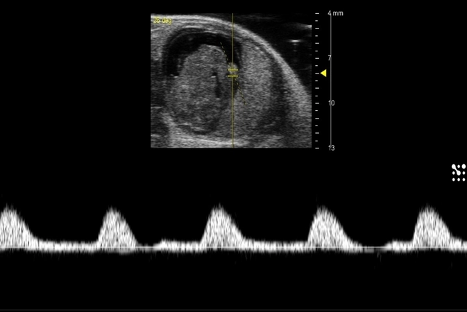
Mouse umbilical artery flow at mid-gestation.
Mouse embryo and placenta showing oxygenation, using photoacoustic imaging.
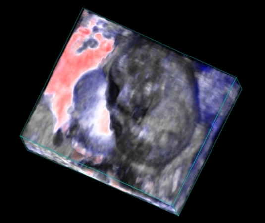
Ultrafast Doppler (UFD) view of a mouse placenta. This was acquired using a UHF29x transducer using VADA (Vevo Advanced Data Acquisition) on the Vevo F2xc imaging platform.
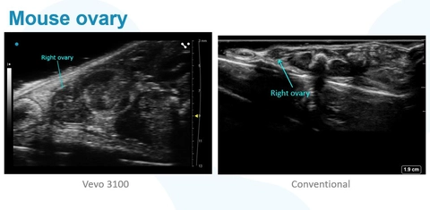
Mouse ovary acquired using ultra high frequency ultrasound (Vevo 3100) against an image of the same mouse ovary using a conventional ultrasound system.
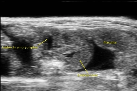
Spina bifida is a neural tube defect that results in incomplete closing of the spinal cord. Spina bifida is associated with abnormalities in the cerebellum and cisterna magna during fetal development. This image acquired using the Vevo 3100.
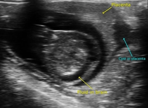
Spina bifida is a neural tube defect that results in incomplete closing of the spinal cord. Spina bifida is associated with abnormalities in the cerebellum and cisterna magna during fetal development. This image acquired using the Vevo 3100.
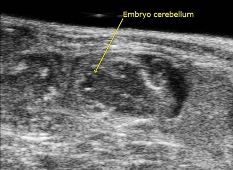
Spina bifida is a neural tube defect that results in incomplete closing of the spinal cord. Spina bifida is associated with abnormalities in the cerebellum and cisterna magna during fetal development. This image acquired using the Vevo 3100.
Spina bifida is a neural tube defect that results in incomplete closing of the spinal cord. Spina bifida is associated with abnormalities in the cerebellum and cisterna magna during fetal development. This image acquired using the Vevo 3100.