
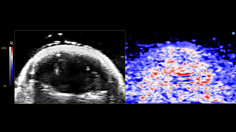
Oxy-Hemo View of Mouse Brain
Oxy-Hemo view of a mouse brain. This was acquired using a UHF29x transducer on the Vevo F2xc imaging platform.
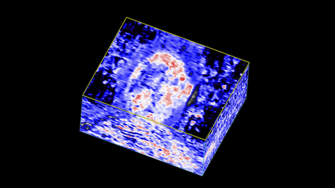
3D Oxy-Hemo View of Mouse Brain
3D Oxy-Hemo view of a mouse brain. This was acquired using a UHF29x transducer on the Vevo F2xc imaging platform.
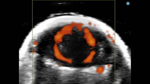
Power Doppler view of Mouse Brain
Power Doppler view of a mouse brain. This was acquired using a UHF29x transducer on the Vevo F2xc imaging platform.
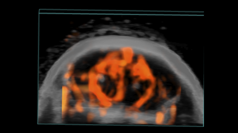
3D Power Doppler view of Mouse Brain
3D Power Doppler view of a mouse brain. This was acquired using a UHF29x transducer on the Vevo F2xc imaging platform.
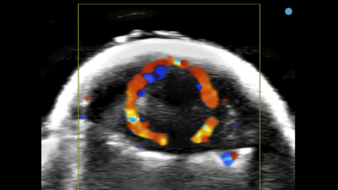
Color Doppler of Mouse Brain
Color Doppler view of a mouse brain. This was acquired using a UHF29x transducer on the Vevo F2xc imaging platform.
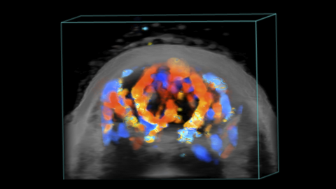
3D Color Doppler of Mouse Brain
3D Color Doppler view of a mouse brain. This was acquired using a UHF29x transducer on the Vevo F2xc imaging platform.
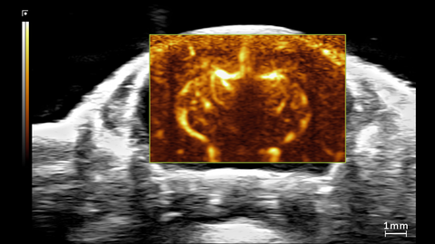
Ultrafast Doppler view of Mouse Brain
Ultrafast Doppler (UFD) view of the brain in a 1.5 month old CD-1 mouse. This was acquired using a UHF29x transducer on the Vevo F2xc imaging platform.
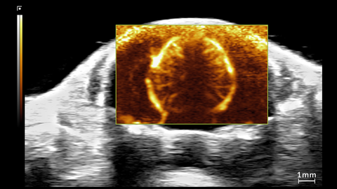
Ultrafast Doppler view of Mouse Brain
Ultrafast Doppler (UFD) view of the brain in a 1.5 month old CD-1 mouse. This was acquired using a UHF29x transducer on the Vevo F2xc imaging platform.
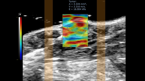
Stiffness map in tumor region
SWE mode overlay showing stiffness map in the tumor region. This was acquired using a UHF57x transducer on the Vevo F2xc imaging platform.
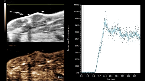
NLC MIP of Mouse Tumor
Maximum intensity projection (MIP) of MicroMarker wash-in acquired in non-linear contrast mode. Contrast region graph shows quantification of the tumor ROI. This was acquired using a UHF57x transducer on the Vevo F2xc imaging platform.
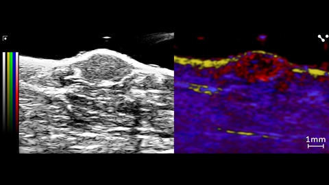
Unmixing from a PA Spectro scan in Mouse Tumor
Unmixing from a PA Spectro scan showing lipid (yellow) in the skin layer, and oxy/deoxyhemoglobin (red/blue) in the tumor and surrounding tissue. Collagen (green) was included in the unmixing, but no noticeable signal above background. This was acquired using a UHF57x transducer on the Vevo F2xc imaging platform.
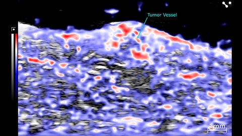
PA Oxyhemo of a Mouse Tumor
PA Oxyhemo scan of a mouse tumor. This was acquired using a UHF57x transducer on the Vevo F2xc imaging platform.
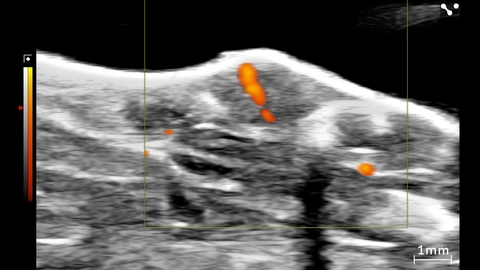
Power Doppler of Tumor in Mouse
Power Doppler of a tumor in a mouse. This was acquired using a UHF57x transducer on the Vevo F2xc imaging platform.

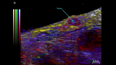
Unmixing from a Spectro scan of a subcutaneous prostate tumor
Unmixing from a Spectro scan of the Subcutaneous prostate tumor and surrounding region. This was acquired using a UHF57x transducer on the Vevo F2xc imaging platform.
Colormap – red/blue = oxy/deoxyhemo; yellow = lipid, green = collagen (theoretical).
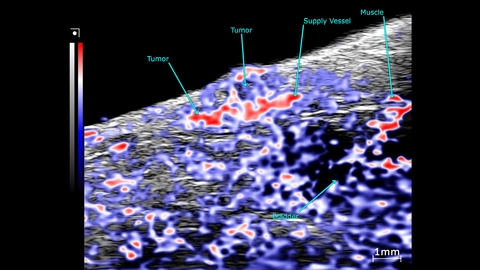
OxyHemo sO2 map of a subcutaneous prostate tumor
PA mode scans showing an OxyHemo sO2 map of a subcutaneous prostate tumor. This image was acquired using a UHF57x transducer on the Vevo F2xc imaging platform.
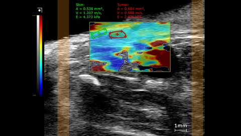
Subcutaneous prostate tumor stiffness map
SWE mode stiffness map showing elastography measurements comparing the tumor to skin inside a mouse. This image was obtained using a UHF29x transducer on a Vevo F2xc imaging platform.