B-Mode
2D Universal Imaging Mode
Visualize and quantify anatomical structures and more
The brightness of each dot is determined by the amplitude of the returned echo signal. This allows for visualization and quantification of anatomical structures, as well as for the visualization of diagnostic and therapeutic procedures for small animal studies.
Images can be exported as still images (TIFF, BMP) or video cine loops (MP4, AVI, GIFF, WMV, DICOM) and can be analyzed with Vevo LAB software.
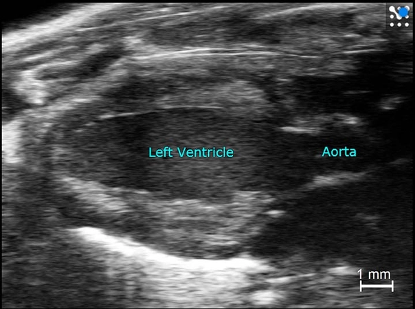
B-Mode Image of a Mouse Heart in Parasternal Long Axis.
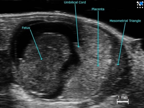
B-Mode Image of a Mid-gestational Mouse.