
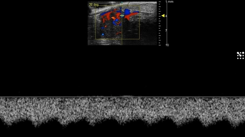
Blood Flow in the Murine Testicular Artery
PW Doppler signal from the testicular artery in a mouse.
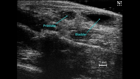
Prostate and Bladder in a Mouse
The prostate and bladder of a male mouse imaged in B-mode and labelled.
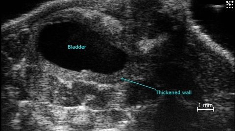
Mouse Bladder with Thickened Walls
Mouse bladder imaged in B-mode with thickened walls labelled.

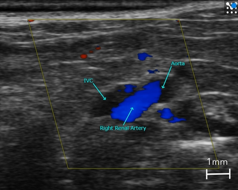
Major Abdominal Blood Vessels
Major abdominal blood vessels in the mouse imaged with color Doppler, including, branching of the right renal artery from the abdominal aorta and the Inferior Vena Cava (IVC).
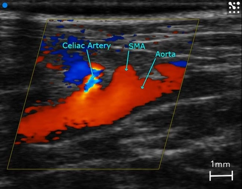
Branching of the Abdominal Aorta
Abdominal aorta in the mouse images with color Doppler, showing branching of the superior mesenteric artery (SMA) and celiac artery.
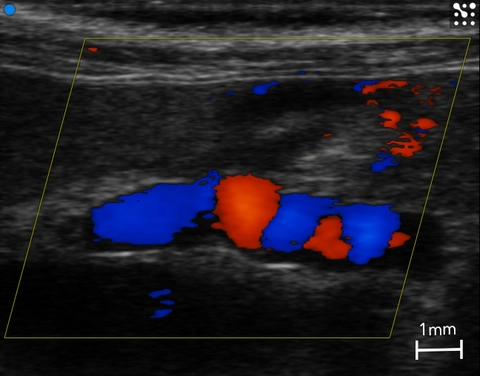
Mouse Portal Vein
Mouse portal vein imaged with color Doppler to show typical swirling blood flow.
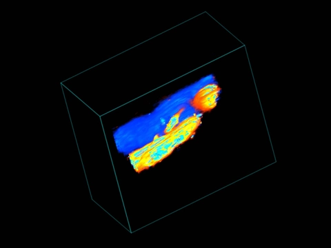
Carotid Fistula
3D Color Doppler image of a carotid fistula in a mouse.
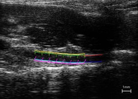
Vascular Strain Analysis in the Abdominal Aorta
Vascular strain analysis on the mouse abdominal aorta using.
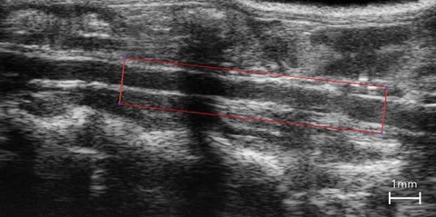
Mouse Abdominal Aorta
Longitudinal section of the abdominal aorta in the mouse to used to measure pulse propagation velocity.
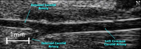
Carotid Artery Bifurcation
B-Mode image of the mouse left common carotid bifurcating into the internal and external carotid arteries.
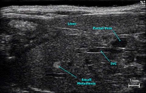
Liver Metastasis
Cross section of the mouse abdomen highlighting major structures, including liver metastasis.
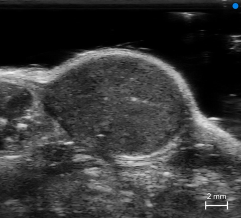
Subcutaneous Tumor
B-Mode image of a subcutaneous tumor in a mouse.
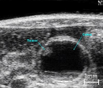
Colon Cancer
B-Mode image of the mouse colon in cross-section with presence of a tumor highlighted.
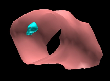
3D Surface Volume of the Mouse Colon with Tumor
Surface view of a 3D volume of a section of mouse colon (in pink) with a tumor (in blue).