


Oxygen saturation in tumor
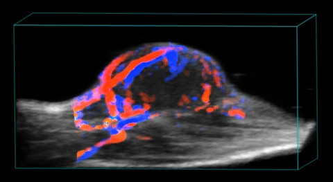
Blood flow in tumor
3D rendered high-resolution ultrasound (greyscale) and color Doppler (orange and blue) image of a subcutaneous tumor showing blood flow.
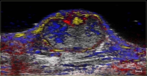
Nanoparticle distribution in tumor
High-resolution ultrasound (greyscale) and spectrally unmixed photoacoustic (color) image of a subcutaneous tumor showing nanoparticle distribution (yellow) as well as oxygenated (red) and deoxygenated (blue) hemoglobin signal.
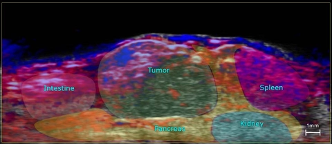
Orthotopic Pancreatic Tumor
High-resolution ultrasound (greyscale) and photoacoustic (red = oxygenated hemoglobin and blue = deoxyhemoglobin) image of an orthotopic pancreatic tumor outlining anatomy.
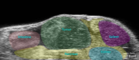
Orthotopic Pancreatic Tumor
High-resolution ultrasound image of an orthotopic pancreatic tumor outlining anatomy.

Blood flow in orthotopic tumor
3D rendered (left) and 2D (right) high-resolution ultrasound (greyscale) and color Doppler (orange and blue) image of an orthotopic pancreatic tumor showing blood flow.
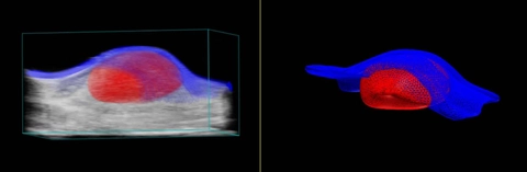
Tumor and Skin Segmentation
3D rendered high-resolution ultrasound image showing wireframe segmentation of a tumor (red) and the overlying skin (blue).
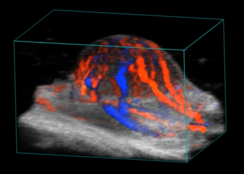
Blood flow in tumor
3D rendered high-resolution ultrasound (greyscale) and color Doppler (orange and blue) image of a subcutaneous tumor showing blood flow.
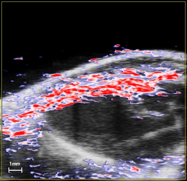
Oxygen saturation in the myocardium
High-resolution ultrasound (greyscale) and photoacoustic (red, white and blue) image of a long axis view of the left ventricle of the mouse heart. The photoacoustic image is a parametric map of oxygen saturation in the myocardium.
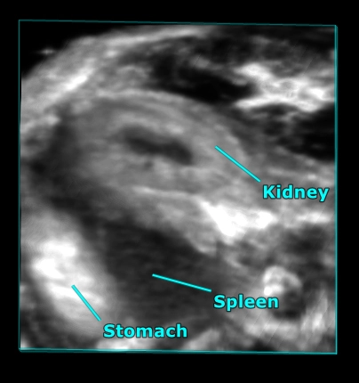
Mouse abdominal organs
3D rendered high-resolution ultrasound image of the mouse abdomen showing the kidney, stomach and spleen.
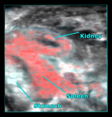
Mouse abdominal organs
3D rendered high-resolution ultrasound (greyscale) and photoacoustic (red) image of the mouse abdomen showing oxygen saturation in the kidney and spleen.
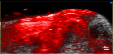
Mouse abdomen showing kidney and spleen
High-resolution ultrasound (greyscale) and photoacoustic (red and white) cross-sectional image of the mouse abdomen showing vasculature in the kidney and spleen.
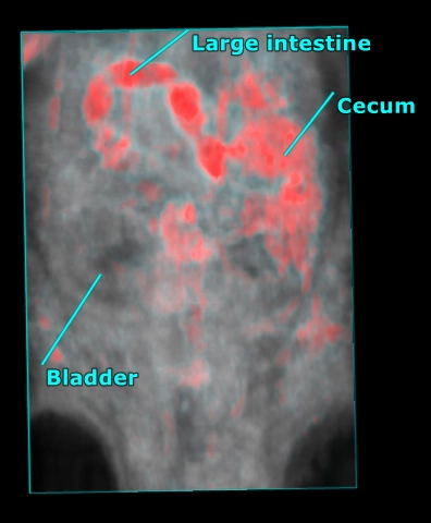
Mouse abdomen
3D rendered high-resolution ultrasound (grey) and photoacoustic (red) image of the whole mouse abdomen showing highly absorbing gut contents overlaid with anatomy as visualized with ultrasound.
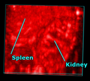
Photoacoustic image of mouse spleen and kidney
3D rendered photoacoustic image of the mouse kidney and spleen.
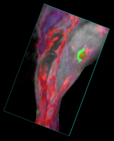
Mouse hindlimb showing blood and ICG
3D rendered high-resolution ultrasound (greyscale) and spectrally unmixed photoacoustic (red, blue and green) image of the mouse hindlbimb. The green color indicates ICG dye in the popliteal lymph node after a hindpaw injection, the red color indicated oxygenated hemoglobin and the blue color indicates deoxygenated hemoglobin.
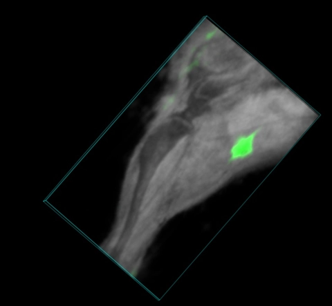
Mouse hindlimb with ICG in lymph node
3D rendered high-resolution ultrasound (greyscale) and spectrally unmixed photoacoustic (green) image of the mouse hindlimb. The green color indicates ICG dye in the popliteal lymph node after a hindpaw injection.
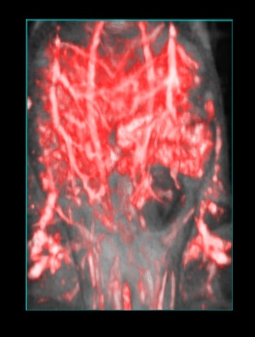
Vasculature and gut in the mouse abdomen
3D rendered photoacoustic (red) and ultrasound (greyscale) image of the mouse abdomen. Vasculature and gut contents can be seen in the photoacoustic image and are overlaid on the anatomical ultrasound image.
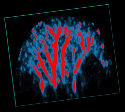
Mouse ear vasculature
3D rendering of a photoacoustic parametric map of oxygen saturation in the mouse ear. Red pixels indicate higher sO2 values.