
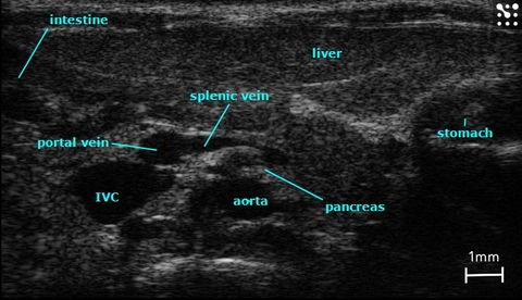
Major Structures in the Mouse Abdomen
B-Mode image of the mouse abdomen with all major structures labelled.
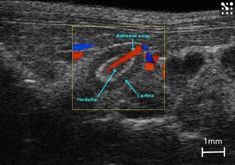
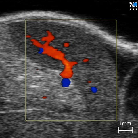
Vasculature in the Mouse Adrenal Gland
Color Doppler image of the mouse adrenal gland with associated vasculature.
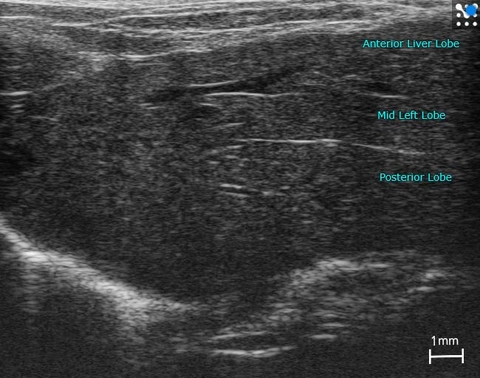
Lobes in the Mouse Liver
Mouse liver with lobes labelled.
Splenic Artery and Vein
Mouse splenic artery and vein with surrounding pancreas imaged in B-Mode.
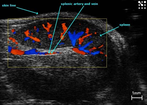
Splenic Vasculature
Color Doppler image of the mouse spleen and splenic vasculature.
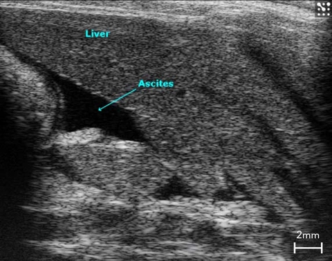
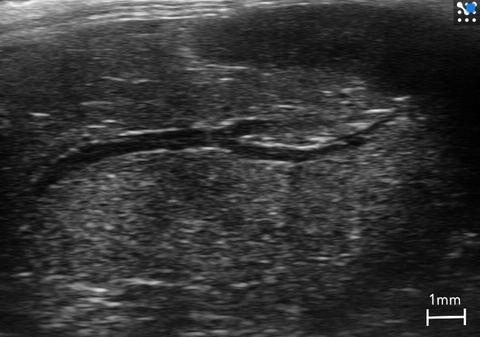
Ascites in the Rat Liver
B-Mode image of ascites in a cirrhotic rat liver.
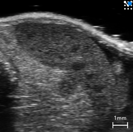
Mouse Spleen
B-Mode image of the spleen in a mouse.
Splenic Vasculature
Color Doppler image of the mouse spleen and splenic vasculature.
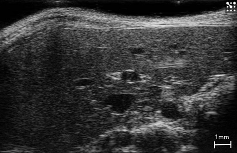
Mouse Liver
Healthy mouse liver imaged in B-Mode.
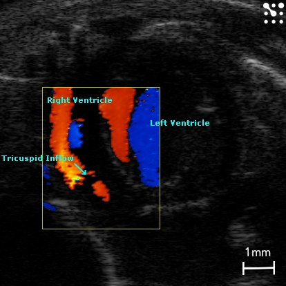
Tricuspid Flow in the Mouse
Apical four chamber view illustrating blood flow through the tricuspid valve, imaged with color Doppler.
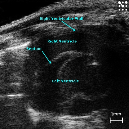
Mouse Right Ventricle
The right ventricle in a mouse imaged in B-Mode.
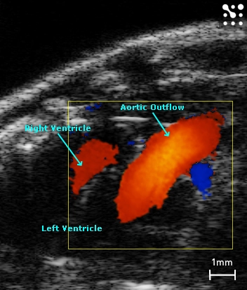
Ascending Aorta from a Parasternal Notch View
Color Doppler image showing blood flow through the ascending aorta and right ventricle from a parasternal notch view in a mouse.
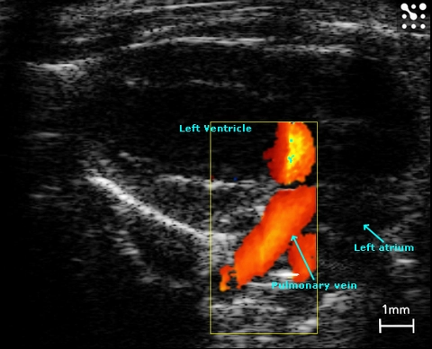
Left Atrium and Pulmonary Veins
Pulmonary veins entering the left atrium in the mouse; imaged with power Doppler.
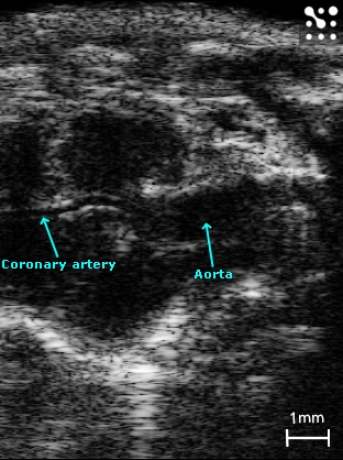
Mouse Coronary Artery
B-Mode image showing the mouse coronary artery branching of the root of the aorta.