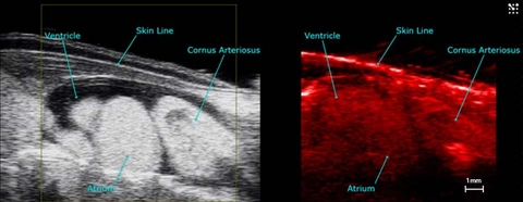
Axolotl heart in PA

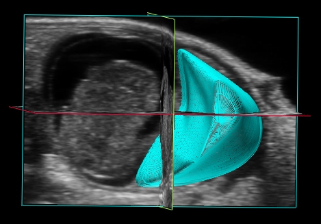
3D Reconstruction of the Placenta
3D reconstruction of the mid-gestational mouse placenta (in blue) with fetus to the left of the image.

Placental Blood Flow
Color Doppler image illustrating blood flow in the fetus, umbilical cord and placenta in a late gestational mouse implantation site.
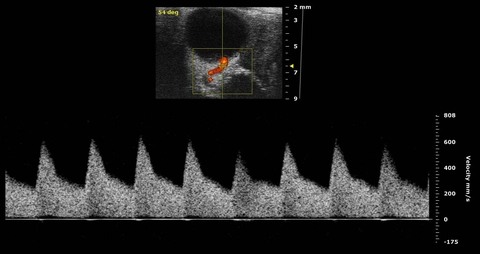
Uterine Artery Blood Flow
PW Doppler of the uterine artery in a mid-gestational mouse.

Umbilical Artery Blood Flow
PW Doppler of the umbilical artery in a mid-gestational mouse.
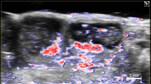
Oxygen Saturation in the Mouse Placenta and Fetus
Oxy-hemo phototacoustics image showing oxygen saturation in two mid-gestational mouse implantation sites. Red indicates oxygenated hemoglobin; blue indicates de-oxygenated hemoglobin.
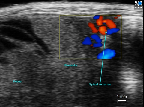
Spiral Arteries in the Mouse Placenta
Color Doppler indicating direction on blood flow in placental spiral arteries in a mid-gestational mouse implantation site.