From Detecting Pregnancy to Measuring Fetal and Placental Oxygenation and Blood Flow
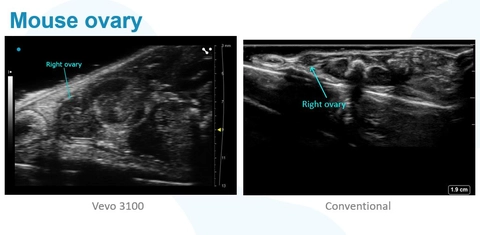
Vevo ultra-high frequency imaging platforms provide superior resolution ideally suited for developmental and pregnancy research in small animals.
The size, structural details and function of the placenta and developing embryo can be measured over multiple gestational time-points without having to sacrifice your animal.
With the Vevo imaging systems you can:
- Detect and date pregnancy with confidence
- Measure size, blood flow and oxygen saturation in the placenta
- Visualize and measure umbilical blood flow
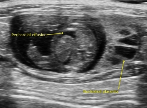
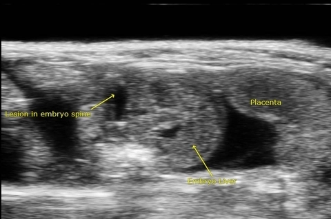
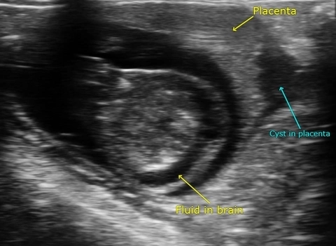
- Visualize and study fetal organ development
- Measure developing heart contractility and blood vessel flow
- Perform precise image-guided embryo injections
Ideal for research on:
- Developmental toxicity and drug trials
- Fetal programming
- Pregnancy research and pregnancy complications
Images of Interest
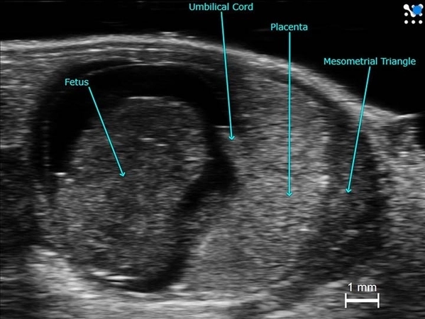
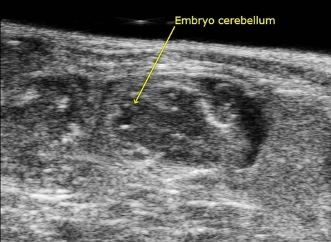
Detect and date pregnancy with confidence
Mouse implantation site at mid-gestation.
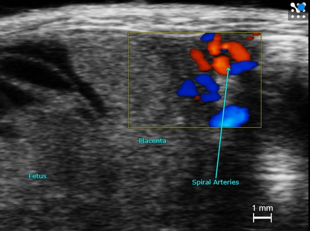
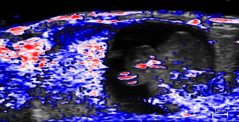
Measure size, blood flow and oxygen saturation
Spiral arteries with Color Doppler in a mid-gestational mouse placenta.
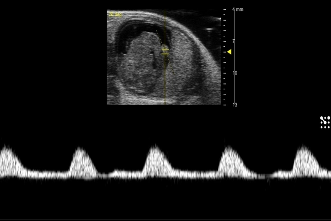
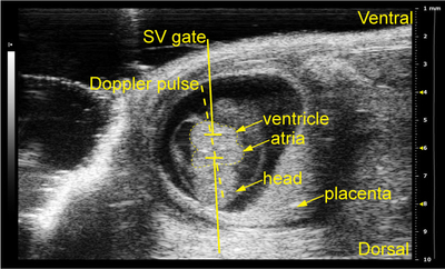
Visualize and measure umbilical blood flow
Mouse umbilical artery flow at mid-gestation.
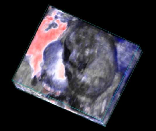
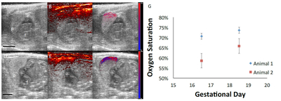
Visualize and study fetal organ development
Mouse embryo and placenta showing oxygenation, using photoacoustic imaging.
Gallery
Publications
TOP PAPER
A Protocol for Evaluating Vital Signs and Maternal-Fetal Parameters Using High-Resolution Ultrasound in Pregnant Mice
STAR Protocols
,
TOP PAPER
Novel insights into the genetic landscape of congenital heart disease with systems genetics
Progress in Pediatric Cardiology
,
TOP PAPER
IL-10 producing B cells rescue mouse fetuses from inflammation-driven fetal death and are able to modulate T cell immune responses
Scientific Reports
,
TOP PAPER
Placenta-specific drug delivery by trophoblast-targeted nanoparticles in mice
Theranostics
, Prenatal hormone stress triggers embryonic cardiac hypertrophy outcome by ubiquitin-dependent degradation of mitochondrial mitofusin 2
iScience
, Phosphodiesterases Mediate the Augmentation of Myogenic Constriction by Inhibitory G Protein Signaling and is Negatively Modulated by the Dual Action of RGS2 and 5
Function
, Maternal exposure to polyethylene micro- and nanoplastics impairs umbilical blood flow but not fetal growth in pregnant mice
Scientific Reports
, Inhibition of Caspase 1 Reduces Blood Pressure, Cytotoxic NK Cells, and Inflammatory T-Helper 17 Cells in Placental Ischemic Rats
International Journal of Molecular Sciences
, Evaluation of recombinant human IGF-1/IGFBP-3 on intraventricular hemorrhage prevention and survival in the preterm rabbit pup model
Scientific Reports
, Request a Quote or Demo
