This recent study by Rutledge et al. compares 4D ultrasound (4D-US) with conventional M-mode and B-mode ultrasound for the assessment of LV function in a myocardial infarct model.
This study by Grune et al. compares LV functional analysis completed using a novel automated 2D-border detection algorithm (referred to as Auto2DE) versus conventional manual 2D echo assessment (2DE).
This study by Heinen et al. compares cardiac functional data in a myocardial infarct model acquired by B-Mode in parasternal long axis (PSLAX) versus Simpson method, versus MRI.
Researchers at the Mouse Imaging Centre of the Hospital for Sick Children in Toronto have established comprehensive methodologies for mouse cardiovascular imaging using high-frequency ultrasound successfully applying the established methodology to the phenotyping of mutant mouse models with human diseases.
Using the Vevo 2100 and ultrasound pulsed-wave (PW) Doppler imaging, Caralynn Wilczewski from Dr. Frank L Conlon's lab has developed a reliable method for performing non-invasive in utero embryonic echocardiography on early gestation mouse embryos. Read more.
This recent article by Platt, et al. uses pulsed-wave Doppler measurement of pulmonary flow (PF) as an alternative method of cardiac output (CO) evaluation in a mouse model of myocardial infarction (MI).
There seems to be some confusion regarding artifacts caused by ribs and the sternum while performing small rodent cardiac exam in the parasternal long axis view. How do you avoid sternum artifacts in your images? Read to find out.
We used Vevo technology to periodically assess left ventricle function during aging and to determine the stage of worst disease condition at which we harvested the heart and performed papillary muscle studies. Read more.
Our new 4D mode on the Vevo 3100 Imaging System will allow you to look at your 3D images over time. Here, a 4D image was taken from a parasternal short axis (SAX) view of the heart.
At the Mouse Imaging Centre of the Hospital for Sick Children in Toronto, we have collaborated with VisualSonics Inc. since early 2000 and used their products from the very first generation to the most advanced versions. Read more.
Dr. Mike Davis' research team at Emory University School of Medicine focuses on various aspects of cardiac regeneration and preservation. His team (part of the joint Biomedical Engineering program at Emory and Georgia Institute of Technology) uses molecular based and biomaterials-based approaches to restore function after cardiac injury. Read more.
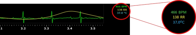
How can you ensure reproducibility in cardiac or contrast imaging? Hypothermia is known to negatively affect the quality of cardiac and contrast reproducibility. The key is monitoring of physiology. Read more.