Neurobiology
Neuroimaging Techniques for Seeing the Mouse Brain in vivo and in Real-time
Visualize functional processes for your small animal neuroscience research including stroke, cancer and in vivo delivery of drugs, stem cells, or other agents.
Compared to other preclinical neuroimaging techniques, Vevo imaging systems are non-invasive, non-ionizing, and offer a more cost effective solution.
Ultra-high frequency ultrasound and photoacoustic imaging is capable of assessing many neurologically relevant parameters in 2D and 3D such as:
- Blood flow
- Perfusion
- Oxygen saturation
- Total Hemoglobin
- Contrast agents (ultrasound microbubbles, NIR dyes, nanoparticles)
In addition, the Vevo BRAIN stereotactic frame and neuroanatomical atlas facilitates mouse brain imaging for increased ease-of-use and reproducibility.
Imaging the Brain
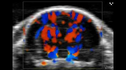
Vasculature in the Brain during Ischemic Stroke
Vasculature in the mouse brain, visualized with color Doppler during transient carotid artery occlusion.
Gallery
Juvenile rat cerebral vasculature

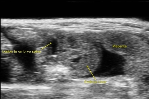
Lesion in Embryo Spine - Spina Bifida Rat Model
Spina bifida is a neural tube defect that results in incomplete closing of the spinal cord. Spina bifida is associated with abnormalities in the cerebellum and cisterna magna during fetal development. This image acquired using the Vevo 3100.
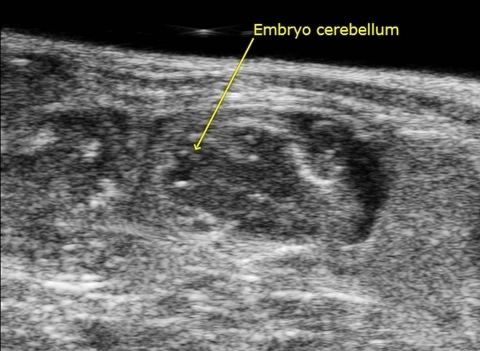
Embryo Cerebellum - Spina Bifida Rat Model
Spina bifida is a neural tube defect that results in incomplete closing of the spinal cord. Spina bifida is associated with abnormalities in the cerebellum and cisterna magna during fetal development. This image acquired using the Vevo 3100.
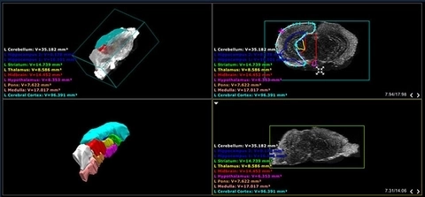
Screenshot of mouse brain atlas
Screenshot of the Vevo LAB software displaying several different views of the CD-1 mouse brain atlas which comes with the Vevo BRAIN stereotactic frame.

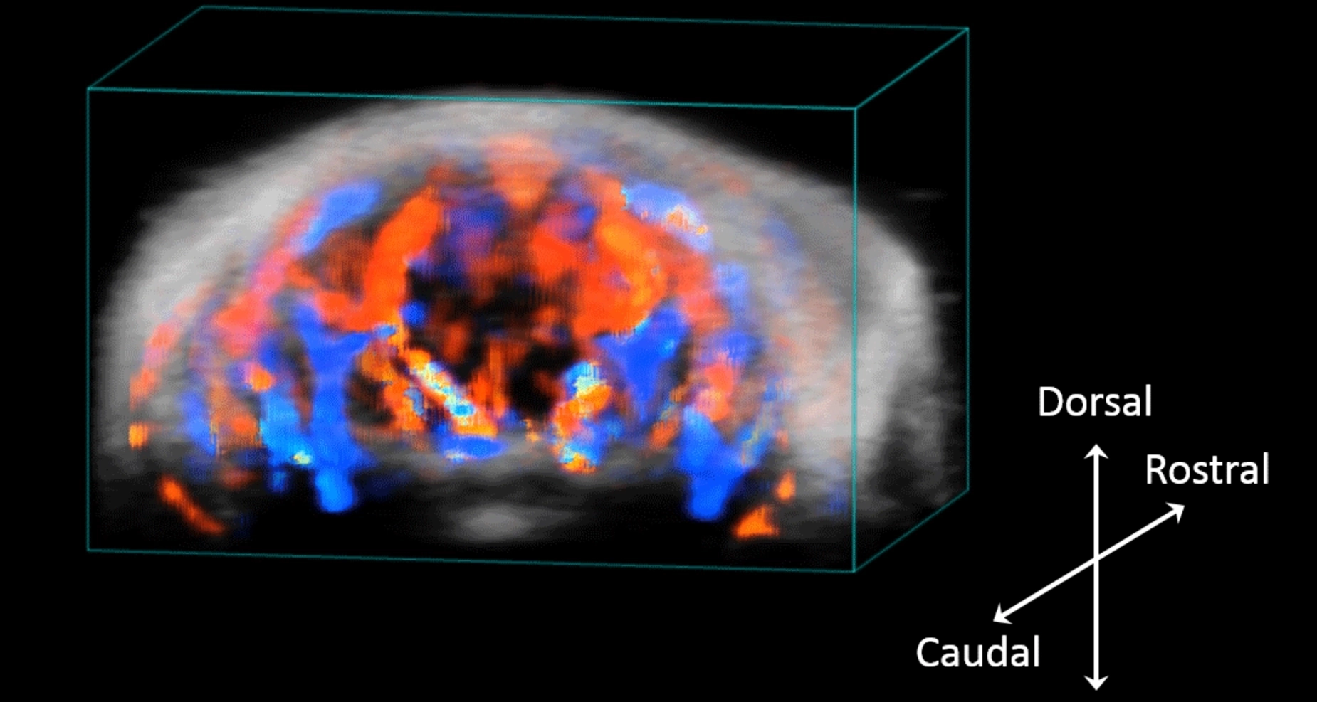
Neuroanatomy of the mouse brain
3D rendered surface view of the mouse brain segmented by anatomical region based on ex vivo high frequency ultrasound images.
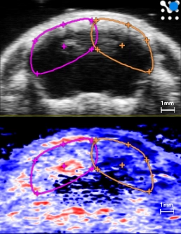
Ischemia/hypoxia stroke model
Plot of oxygen saturation (sO2) over time in the left (pink) and right (orange) cortical and subcortical hemisphere ROIs while restoring breathed oxygen to 100% from 7.5% post ischemia/hypoxia for stroke induction. Ultrasound (top) and photoacoustic (bottom) coronal images in the right panel show a frame of mouse brain post ischemia/hypoxia.
Publications
TOP PAPER
In vivo imaging in experimental spinal cord injury – Techniques and trends
Brain and Spine
,
TOP PAPER
Early cerebrovascular and long-term neurological modifications ensue following juvenile mild traumatic brain injury in male mice
Neurobiology of Disease
,
TOP PAPER
Transcranial Photoacoustic Detection of Blood-Brain Barrier Disruption Following Focused Ultrasound-Mediated Nanoparticle Delivery
Molecular Imaging and Biology
,
TOP PAPER
Multimodal and multiscale optical imaging of nanomedicine delivery across the blood-brain barrier upon sonopermeation
Theranostics
,
TOP PAPER
Exploration of melanoma metastases in mice brains using endogenous contrast photoacoustic imaging
International Journal of Pharmaceutics
, Micro-ultrasound based characterization of cerebrovasculature following focal ischemic stroke and upon short-term rehabilitation
Journal of Cerebral Blood Flow and Metabolism
, Evaluation of recombinant human IGF-1/IGFBP-3 on intraventricular hemorrhage prevention and survival in the preterm rabbit pup model
Scientific Reports
, Corticospinal-specific Shh overexpression in combination with rehabilitation promotes CST axonal sprouting and skilled motor functional recovery after ischemic stroke
Molecular Neurobiology
, Transplantation of active nucleus pulposus cells with a keep-charging hydrogel microsphere system to rescue intervertebral disc degeneration
Journal of Nanobiotechnology
, 

Request a Quote or Demo