Cardiology
Preclinical Echocardiography for Cardiology Research
Ultra-high frequency (UHF) ultrasound provides superior resolution and will have an incredible impact on cardiovascular research in your animal models.
Perform reliable echocardiography in any animal model for translational cardiovascular research with in vivo resolution down to 30 microns.
Vevo ultrasound imaging platforms provide the best temporal and spatial resolution compared to MRI, conventional ultrasound and CT. The Vevo F2 system can perform preclinical echocardiography in animal models all the way from zebra fish and mice to pigs. Vevo technology has been established as a gold standard technology for mouse cardiac imaging, having been utilized in over 3,000 peer-reviewed publications.
With the Vevo imaging systems you can:
- Perform cardiovascular phenotyping for cardiac structure and function, ideal for stress echo and cardiotoxicity studies
- Image the heart in 4D (with Color or Power Doppler integration)
- Quantify diastolic dysfunction
- Evaluate early signs of cardiac dysfunction with cardiac strain (global or regional synchrony and deformation)
- Perform image-guided cardiac injections
- Monitor full animal physiology while imaging, including body temperature, ECG and respiratory rate
- Assess myocardial oxygen saturation
Cardiac 4D Imaging
Quantify ejection fraction and fractional shortening with more accuracy than traditional echocardiography, and faster than cardiac MRI!
Cardiovascular Workshop
The Heart of the Matter
If you missed this advanced workshop on COVID-19 and other preclinical cardiac disease models, here's your chance to catch up.
Gallery
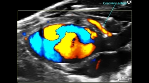
Cardiac Flow Dynamics and Coronary Artery
Cardiac flow dynamics and Coronary Artery visualized with Color Doppler EKV on the Vevo F2 System.
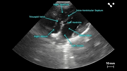
Apical 4 Chamber View of a Beagle
Apical 4 chamber view of a beagle scanned using a P5-1 transducers on the Vevo F2. Images courtesy of Drs. Kenneth Hoyt and Jay Griffin at Texas A&M University.


Color Doppler of Mouse Aortic Arch
Color Doppler image of an aortic arch in a female mouse, acquired using a UHF57x transducer on the Vevo F2.
Publications
TOP PAPER
Endothelial cells drive organ fibrosis in mice by inducing expression of the transcription factor SOX9
Science Translational Medicine
,
TOP PAPER
P2Y12 Inhibition in Murine Myocarditis Results in Reduced Platelet Infiltration and Preserved Ejection Fraction
Cells
,
TOP PAPER
Embryonic echocardiography for assessment of congenital and functional cardiac defects
STAR Protocols
,
TOP PAPER
Quantification of murine myocardial infarct size using 2-D and 4-D high-frequency ultrasound
American Journal of Physiology-Heart and Circulatory Physiology
,
TOP PAPER
Improving characterization of hypertrophy-induced murine cardiac dysfunction using four-dimensional ultrasound derived strain mapping
American Journal of Physiology-Heart and Circulatory Physiology
,
TOP PAPER
Machine learning driven contouring of high-frequency four-dimensional cardiac ultrasound data
Applied Sciences (Switzerland)
,
TOP PAPER
Induced pluripotent stem cell intervention rescues ventricular wall motion disparity, achieving biological cardiac resynchronization post-infarction
Journal of Physiology
, Resources

Request a Quote or Demo