
Cardiovascular Biology Gallery

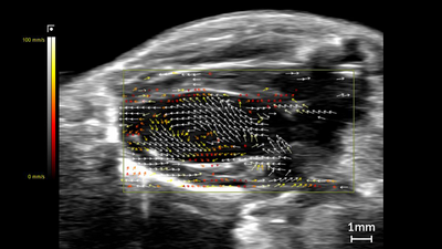
Mouse heart (Vector Flow Imaging)
Vector Flow Imaging (beta feature) shown in a mouse heart. Scanned using a UHF29x transducer on the Vevo F2xc platform.
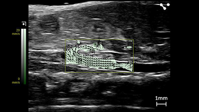
Mouse Abdominal Aorta (Vector Flow Imaging)
Vector Flow Imaging (beta feature) showing the mouse abdominal aorta. Scanned using a UHF29x transducer on the Vevo F2xc platform.
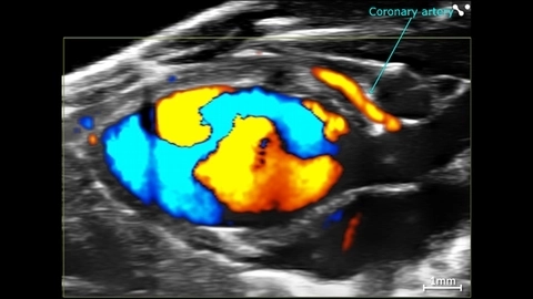
Cardiac Flow Dynamics and Coronary Artery
Cardiac flow dynamics and Coronary Artery visualized with Color Doppler EKV on the Vevo F2 System.
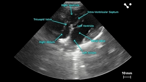
Apical 4 Chamber View of a Beagle
Apical 4 chamber view of a beagle scanned using a P5-1 transducers on the Vevo F2. Images courtesy of Drs. Kenneth Hoyt and Jay Griffin at Texas A&M University.
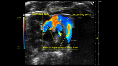
Color Doppler of Mouse Aortic Arch
Color Doppler image of an aortic arch in a female mouse, acquired using a UHF57x transducer on the Vevo F2.
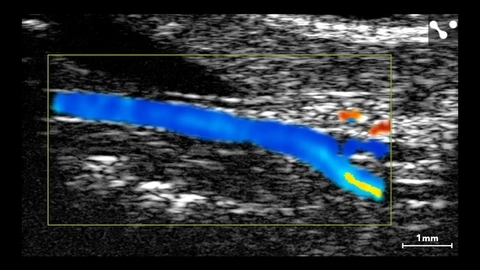
Murine Carotid Artery
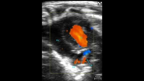
Murine Apical Color Doppler
Murine apical color Doppler scanned using a UHF46x transducer.
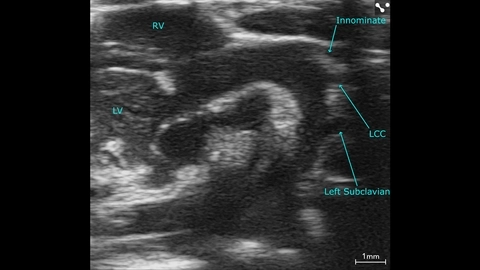
Mouse Aortic Arch
Mouse aortic arch scanned using a UHF57x transducer.
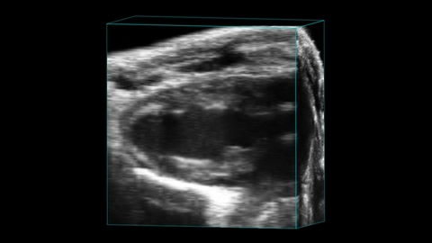
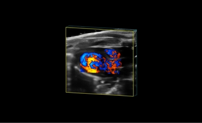
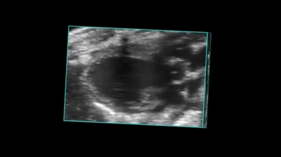
Parasternal Long Axis of Mouse Heart in 4D
Parasternal long axis of a mouse heart in 4D scanned using the UHF46x transducer.

Mitral flow in a small dog