
Ultrafast Doppler Gallery
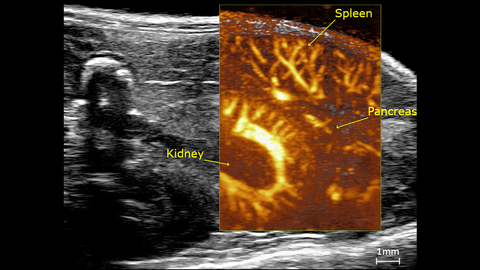
Ultrafast Doppler: Mouse Abdominal
The mouse abdominal ultrasound image showing the vasculature of the pancreas, spleen and the kidney using Ultrafast Doppler. Acquired using a UHF46x transducer on a Vevo F2xc ultrasound system.
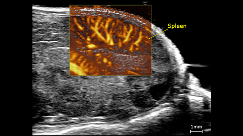
Ultrafast Doppler: Spleen Vasculature
The mouse abdominal ultrasound image showing the spleen vasculature using Ultrafast Doppler. Acquired using a UHF46x transducer on a Vevo F2xc ultrasound system.
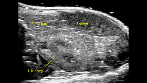
Ultrafast Doppler: Pancreas, Spleen and Left Kidney
The mouse abdominal ultrasound image showing the pancreas, spleen and the left kidney using Ultrafast Doppler. Acquired using a UHF46x transducer on a Vevo F2xc ultrasound system.

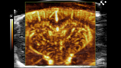
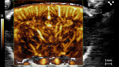
Rat Brain - Cranial Window: Ultrafast Doppler Mode
Ultrafast Doppler (UFD) showing the rat brain cranial window; scanned using a UHF29x transducer on the Vevo F2xc platform.
Images collected courtesy of BioMaps laboratory (Paris-Saclay University), Lim Anna Sieu, Anthony Novell, Jean-Luc Gennisson.
Rat Brain - Cranial Window: Ultrafast Doppler Mode
Ultrafast Doppler (UFD) showing the rat brain cranial window; scanned using a UHF29x transducer on the Vevo F2xc platform.
Images collected courtesy of BioMaps laboratory (Paris-Saclay University), Lim Anna Sieu, Anthony Novell, Jean-Luc Gennisson.
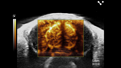
Ultrafast Doppler Mode (UFD) in the Brain with Bubbles
Ultrafast Doppler Mode (UFD) showing the mouse brain transcranial with max bubbles; scanned using a UHF29x transducer on the Vevo F2xc platform.
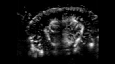
Ultrafast Doppler Mode (UFD) in the Brain
Ultrafast Doppler (UFD) showing the mouse brain transcranial, scanned using a UHF29x transducer on the Vevo F2xc platform.
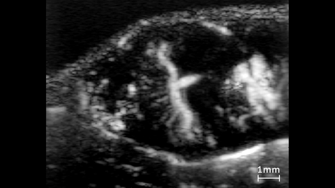
Ultrafast Doppler view of Placenta
Ultrafast Doppler (UFD) view of a mouse placenta. This was acquired using a UHF29x transducer using VADA (Vevo Advanced Data Acquisition) on the Vevo F2xc imaging platform.
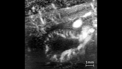
Ultrafast Doppler view of Kidney
Ultrafast Doppler (UFD) view of a mouse kidney. This was acquired using a UHF29x transducer using VADA (Vevo Advanced Data Acquisition) on the Vevo F2xc imaging platform.
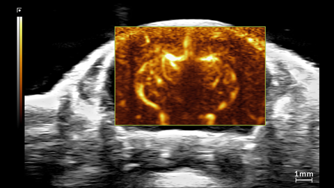
Ultrafast Doppler view of Mouse Brain
Ultrafast Doppler (UFD) view of the brain in a 1.5 month old CD-1 mouse. This was acquired using a UHF29x transducer on the Vevo F2xc imaging platform.
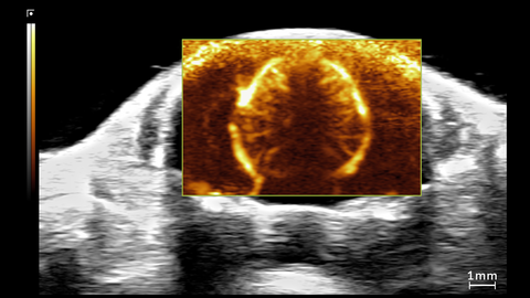
Ultrafast Doppler view of Mouse Brain
Ultrafast Doppler (UFD) view of the brain in a 1.5 month old CD-1 mouse. This was acquired using a UHF29x transducer on the Vevo F2xc imaging platform.