
Vevo LAZR-X20 Gallery

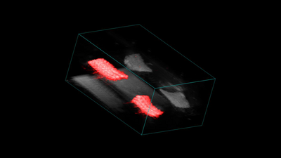
3D Render of Hernia Meshes
3D render of overlaid ultrasound and photoacoustic signal from 2 labelled and 2 control hernia meshes.
Courtesy of Marie Z. Cilliers, Patrick Lab, Centre for Advanced Biomedical Imaging, UCL
Interested in learning more? Read the paper here: Noninvasive Photoacoustic Imaging of Microscale Material Degradation in a Poly(4-hydroxybutyrate) Resorbable Hernia Mesh | ACS Applied Polymer Materials
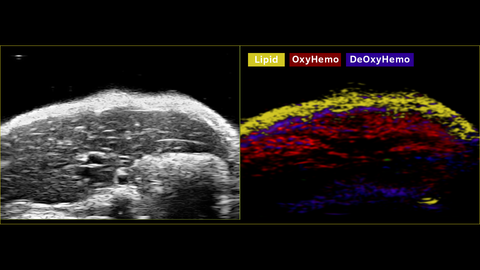
Multiwavelength PA Imaging - Mouse Liver
Multi-wavelength photoacoustic imaging of a mouse liver. Lipid in yellow, oxyhemoglobin in red, and deoxyhemoglobin in blue. This was acquired using a UHF29x transducer on the Vevo F2xc imaging platform.
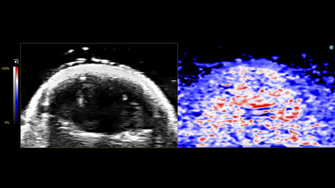
Oxy-Hemo View of Mouse Brain
Oxy-Hemo view of a mouse brain. This was acquired using a UHF29x transducer on the Vevo F2xc imaging platform.
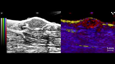
Unmixing from a PA Spectro scan in Mouse Tumor
Unmixing from a PA Spectro scan showing lipid (yellow) in the skin layer, and oxy/deoxyhemoglobin (red/blue) in the tumor and surrounding tissue. Collagen (green) was included in the unmixing, but no noticeable signal above background. This was acquired using a UHF57x transducer on the Vevo F2xc imaging platform.
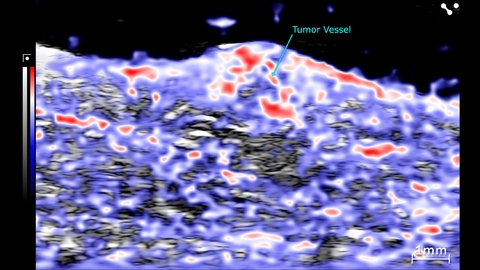
PA Oxyhemo of a Mouse Tumor
PA Oxyhemo scan of a mouse tumor. This was acquired using a UHF57x transducer on the Vevo F2xc imaging platform.
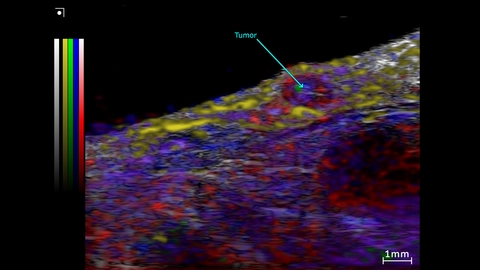
Unmixing from a Spectro scan of a subcutaneous prostate tumor
Unmixing from a Spectro scan of the Subcutaneous prostate tumor and surrounding region. This was acquired using a UHF57x transducer on the Vevo F2xc imaging platform.
Colormap – red/blue = oxy/deoxyhemo; yellow = lipid, green = collagen (theoretical).
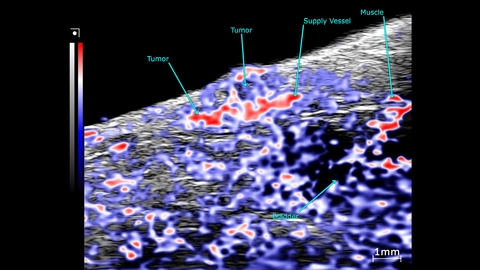
OxyHemo sO2 map of a subcutaneous prostate tumor
PA mode scans showing an OxyHemo sO2 map of a subcutaneous prostate tumor. This image was acquired using a UHF57x transducer on the Vevo F2xc imaging platform.
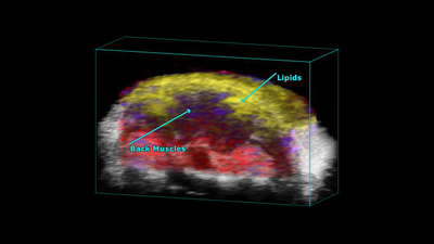
Mouse Scapular
PA-Mode 3D unmixing of a mouse scapular, highlighting the back muscles and lipids. Captured using a UHF29x transducer.
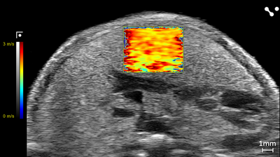
Shear Wave Elastography: NAFLD/MASLD Mouse - Velocity
Liver in a NAFLD/MASLD Mouse showing Velocity (m/s) through Shear Wave Elastography scanned using a UHF29x transducers on a Vevo F2 system.
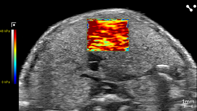
Shear Wave Elastography: NAFLD/MASLD Mouse - Stiffness
Liver in a NAFLD/MASLD Mouse showing Stiffness (kPa) through Shear Wave Elastography scanned using a UHF29x transducers on a Vevo F2 system.
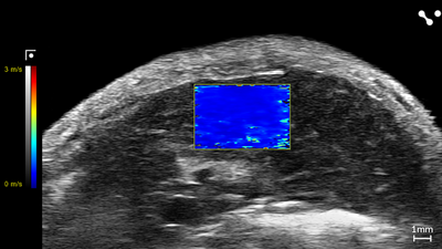
Shear Wave Elastography: Control Mouse - Velocity
Liver in a Control Mouse showing Velocity (m/s) through Shear Wave Elastography scanned using a UHF29x transducers on a Vevo F2 system.
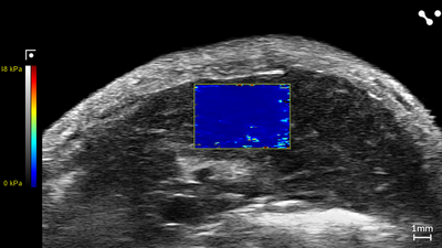
Shear Wave Elastography: Control Mouse - Stiffness
Liver in a Control Mouse showing Stiffness (kPa) through Shear Wave Elastography scanned using a UHF29x transducers on a Vevo F2 system.
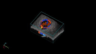
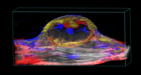
Nanoparticle distribution in tumor
3D rendered high-resolution ultrasound (greyscale) and spectrally unmixed photoacoustic (red, blue and gold) image of a subcutaneous tumor showing nanoparticle distribution (yellow) as well as oxygenated (red) and deoxygenated (blue) hemoglobin signal.
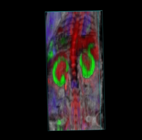
Biodistribution of photoacoustic contrast agent and signals from oxy and deoxyhemoglobin
Whole body biodistribution of photoacoustic contrast agent Angiostamp800TM (green), along with signals from oxy (red) and deoxyhemoglobin (blue).