Vascular Biology
Preclinical Ultrasound for Vascular Biology Research
Visualize and Quantify Vascular Function
Ultra-high Frequency (UHF) Vevo imaging systems allow for non-invasive, real-time evaluation of vascular function with resolution down to 30 microns.
With UHF imaging and software, you can:
- Quantify pulse wave velocity and vascular resistance
- Detect aneurysm and plaque formation with 3D reconstruction
- Analyze vessel wall characteristics (i.e. diameter, strain, velocity, stiffness, etc.)
- Quantify blood flow and perfusion
Perfect for Animal Models of:
- Atherosclerosis
- Aneurysm
- Arteriosclerosis
- Vascular Ischemia
Vascular Biology
Right Femoral Artery Bifurcation in a Mouse
Gallery
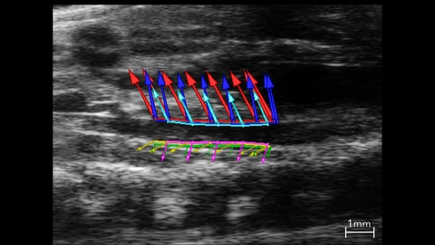
Abdominal Aorta Used In Strain Analysis
B-mode image of the abdominal aorta with vessel wall velocity vector arrows used in strain analysis.
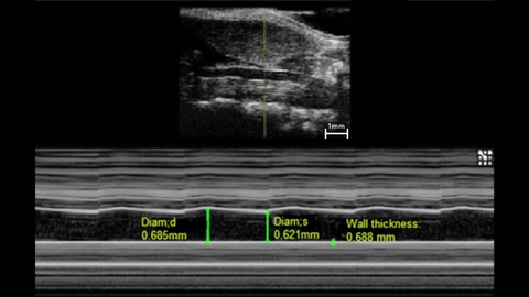
M-Mode of a Mouse Carotid Artery
M-mode image of mouse left common carotid.
Measurements: wall thickness and vessel diameter in systole and diastole.
Splenic Vasculature in a Mouse
Murine Renal Vasculature in 3D
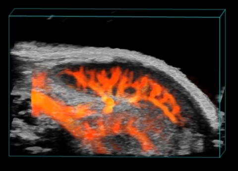
3D Spleen and Kidney Vasculature
3D Power Doppler rendering of a mouse spleen and kidney, highlighting splenic and renal vasculature.

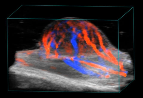
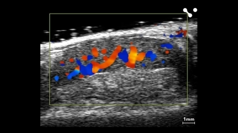
Blood flow in tumor
3D rendered high-resolution ultrasound (greyscale) and color Doppler (orange and blue) image of a subcutaneous tumor showing blood flow.
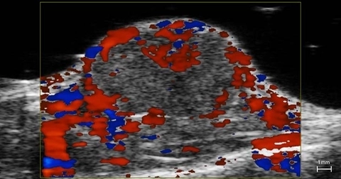
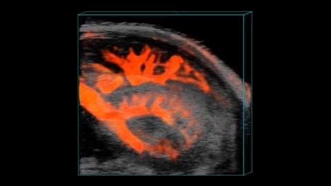
Blood flow in tumor
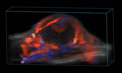
Blood flow in tumor
3D rendered high-resolution ultrasound (greyscale) and color Doppler (orange and blue) image of a subcutaneous tumor showing blood flow.
Publications
TOP PAPER
Fast super-resolution ultrasound microvessel imaging using spatiotemporal data with deep fully convolutional neural network
Physics in Medicine and Biology
,
TOP PAPER
A Protocol for Evaluating Vital Signs and Maternal-Fetal Parameters Using High-Resolution Ultrasound in Pregnant Mice
STAR Protocols
,
TOP PAPER
Scutellarin Prevents Angiogenesis in Diabetic Retinopathy by Downregulating VEGF/ERK/FAK/Src Pathway Signaling
Journal of Diabetes Research
,
TOP PAPER
Pharmacological inhibition of Notch signaling regresses pre-established abdominal aortic aneurysm
Scientific Reports
,
TOP PAPER
Strain Mapping From Four-Dimensional Ultrasound Reveals Complex Remodeling in Dissecting Murine Abdominal Aortic Aneurysms
Journal of Biomechanical Engineering
,
TOP PAPER
Strain mapping from 4D ultrasound reveals complex remodeling in dissecting murine abdominal aortic aneurysms.
Journal of biomechanical engineering
,
TOP PAPER
Development and growth trends in angiotensin II-induced murine dissecting abdominal aortic aneurysms
Physiological Reports
,
TOP PAPER
Impairment of an Endothelial NAD + -H 2 S Signaling Network Is a Reversible Cause of Vascular Aging
Cell
,
TOP PAPER
A critical role for macrophages in neovessel formation and the development of stenosis in tissue-engineered vascular grafts
The FASEB Journal
, Request a Quote or Demo