Urogenital
A Simple and Accurate Imaging Solution for Bladder and Prostate Cancer Research

Mouse bladder with thickened walls.
Use Vevo systems to perform:
- Volumetric analysis
- Vascular assessment
- Image-guided cell injection to establish orthotopic tumor models
- Wall thickness measurements (i.e. bladder)
Ultra-high frequency ultrasound for urogenital imaging is ideal for:
- Cancer research – prostate, ovarian, bladder, testicular
- Endometriosis
- Assisted Reproductive Technologies
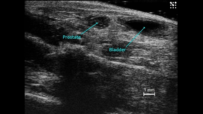
Mouse prostate in B-mode.
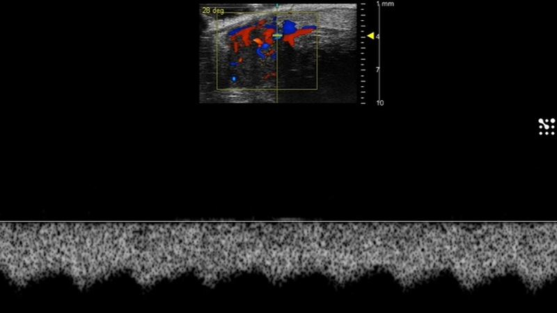
Mouse testicular artery blood flow.
Gallery


Ovary, spleen and kidney
B-mode image of the ovary, spleen and kidney, acquired using a UHF57x transducer on the Vevo F2.

Mouse ovary and kidney
B-mode image of the mouse ovary and kidney, acquired using a UHF57x transducer on the Vevo F2.
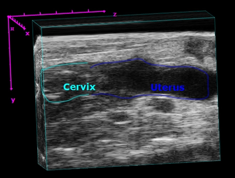
Mouse Cervix and Uterus
3D visualization of the mouse cervix and uterus from a sagittal view.
Publications
TOP PAPER
A simple and robust nanosystem for photoacoustic imaging of bladder cancer based on α5β1-targeted gold nanorods
Journal of Nanobiotechnology
,
TOP PAPER
Early diagnosis of bladder cancer by photoacoustic imaging of tumor-targeted gold nanorods
Photoacoustics
,
TOP PAPER
Evaluation of ductal carcinoma in situ grade via triple-modal molecular imaging of B7-H3 expression
npj Breast Cancer
, Spectral photoacoustic imaging of age-related reproductive tract collagen changes in a mouse model of prolapse
Photoacoustics
, Tumor necrosis factor alpha inhibition improves fetal growth in a rat model of preeclampsia
Placenta
, In vivo photoacoustic imaging of swine ureters injected with methylene blue
Journal of Biomedical Optics
, Autophagy Plays a Suppressive Role in Bladder Tumor Formation in an Orthotopic Mouse Model and Bladder Cancer Patient Specimens
Kaohsiung Journal of Medical Sciences
, Shifting the paradigm of PSMA delivery in prostate cancer for internal radiotherapy: An innovative ultrasound-mediated approach
Biomedicine & Pharmacotherapy
, IL-33 Signaling Inhibition Leads to a Preeclampsia-Like Phenotype in Pregnant Rats
American Journal of Reproductive Immunology
, Request a Quote or Demo