Oncology
Your Powerful Solution for Cancer Research
Cancer Imaging for Translational Research
Characterize tumor tissue in vivo, non-invasively, detect lesions early, monitor tumor development and assess response to therapy!
Vevo for Oncology: A Longitudinal Study Workflow
For more information on the longitudinal study workflow, download our Vevo for Oncology document.
With Vevo cancer imaging technology for translational research, you can perform image-guided injections to create orthotopic tumors, conduct high-throughput screenings for tumor growth and detect and monitor growth and response of tumors to therapy at multiple time points in preclinical disease models all in vivo. Vevo technology allows you to assess tumor size and volume accurately. Add imaging the tumor microenvironment for changes in angiogenesis, hypoxia, molecular markers, and drug delivery and you have a powerful multi-modal platform for oncology research.
With the Vevo Imaging Systems you can:
- Screen for very small lesions (resolution down to 30µm)
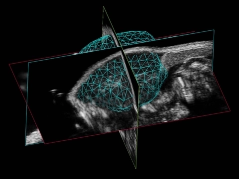
- Accurately quantify volume of orthotopic and subcutaneous tumors longitudinally
- Visualize and quantify angiogenesis, vasculature and perfusion
- Measure hypoxia
- Assess biomarker or drug distribution
- Develop models using ultrasound-guided injections


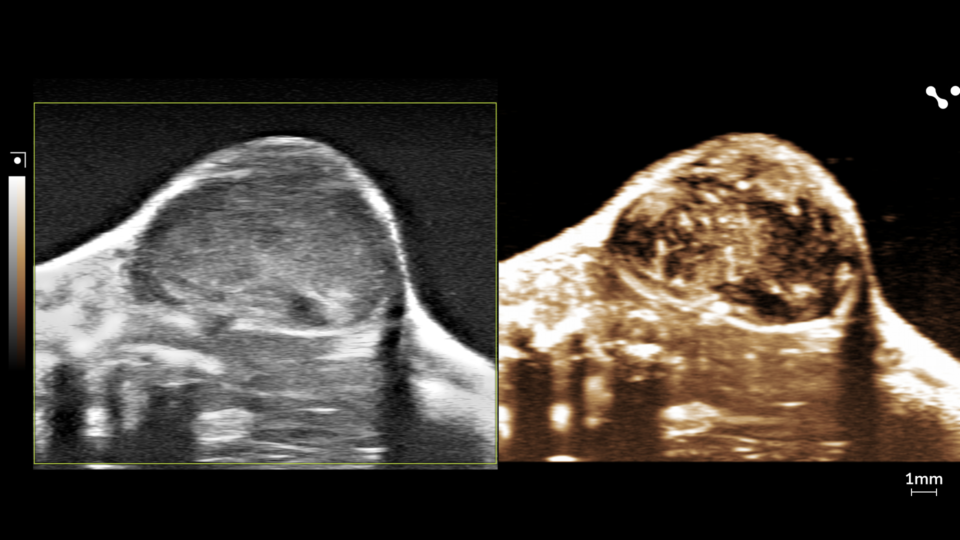
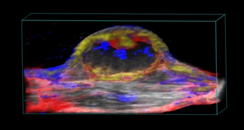
Oxygen saturation in a mouse subcutaneous tumor imaged using photoacoustics.
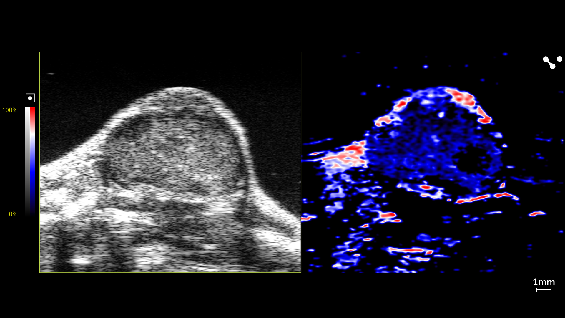
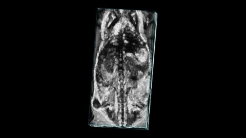
Bolus injection of non-targeted contrast agent to look at perfusion kinetics in a mouse bladder tumor.
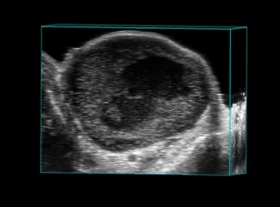
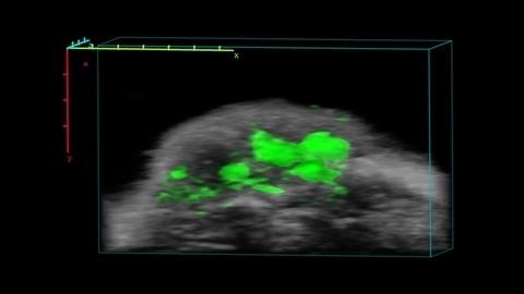
(Left) B-Mode image-guided injection into a mouse tumor with injection and aspiration. This technique can be used for cancer model development.
Gain insights into various tumor models including:
- Orthotopic pancreatic tumors
- Breast tumors
- Prostate and ovarian cancer
- Bladder tumors
- Gliomas
- Patient-dervied xenografts
View literature published using the Vevo systems for these models and others.
See images and videos related to Cancer Imaging for Translational Research.
Learn more about Cancer Imaging for Translational Research by attending one of our courses, webinars or events
Downloaded or access resources related to Cancer Imaging for Translational Research
Light-assisted hierarchical intratumoral penetration and programmed antitumor therapy based on tumor microenvironment (TME)-amendatory and self-adaptive polymeric nanoclusters
Generation of Pancreatic Organoid-Derived Isografts
Gambogic acid augments black phosphorus quantum dots (BPQDs)-based synergistic chemo-photothermal therapy through downregulating heat shock protein expression
Nanostructural Control Enables Optimized Photoacoustic–Fluorescence–Magnetic Resonance Multimodal Imaging and Photothermal Therapy of Brain Tumor
Sequential delivery of nanoformulated α-mangostin and triptolide overcomes permeation obstacles and improves therapeutic effects in pancreatic cancer
Thank you for your patience. Your search results are loading.
Essential Links
Request a Quote or Demo
Contact UsWEBINAR:
Assessment of Murine Colorectal Cancer by UHF Ultrasound using 3D Reconstruction and Non-Linear Contrast Imaging
“Utilizing the Vevo2100 to assess orthotopic colorectal cancer in mice has proven instrumental to our data quality. Instead of using endpoint-only data for our studies, we are now able to collect high-resolution longitudinal data, making sure to capture responses to treatment that otherwise would have been missed. The true value lies in the fact that we can collect data that is giving us tumor VOLUMES rather than just tumor areas, enabling us to know more about the tumor morphology. And using non-linear contrast combined with microbubbles we are able to further investigate the vascularity of the colon as a whole as well as individual tumors. Knowing how those tumors recruit vessels to feed them and if our treatments can influence this vascular recruitment may be the key to finding worthwhile therapies.”
~ Jessical Freeling, MS, VT, LATG, Physiology Core Laboratory Manager,
The University of South Dakota