Oncology Gallery
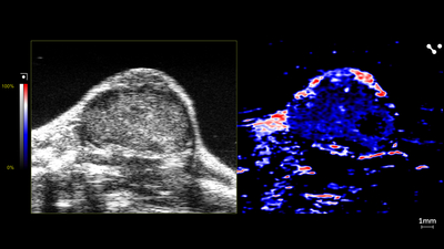
Oyx-hemo Imaging of Mouse Tumor

Oxy-hemo image of a mouse tumor showing shear wave elastography, scanned using a UHF29x transducer.

Contrast Imaging of Mouse Tumor

Contrast image of a mouse tumor showing shear wave elastography, scanned using a UHF29x transducer.
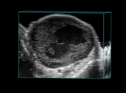
Breast tumor in 3D

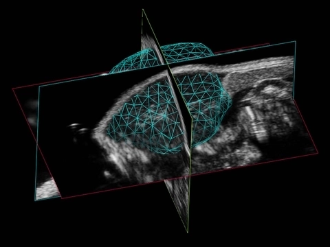
Tumor 3D

3D Volume Reconstruction of a Murine Tumor
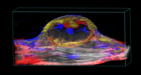
Nanoparticle distribution in tumor

3D rendered high-resolution ultrasound (greyscale) and spectrally unmixed photoacoustic (red, blue and gold) image of a subcutaneous tumor showing nanoparticle distribution (yellow) as well as oxygenated (red) and deoxygenated (blue) hemoglobin signal.
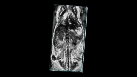
Mouse whole body with tumor

Whole body image of a mouse with a subcutaneous tumor visible on the flank, imaged with high frequency ultrasound on the Vevo F2 system.
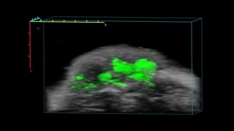
ICG localization in tumor


Ultrasound guided free hand injection into the murine pancreas
Acquired using the Vevo 3100.

40uL volume injected into the mouse pancreas transcutaneously with image-guided injection
Acquired using the Vevo 3100.

Parametric Map of Oxygen Saturation in an Orthotopic Breast Tumor Model
In this parametric map of oxygen saturation in an orthotopic breast tumor, the breathed oxygen concentration is varied from 100% to 5% back to 100% and the resulting changes in sO2 can be observed in different areas.
Note that the sO2 in the tumor interior does not change appreciably during the breathed oxygen challenge, indicating impaired function of the vasculature there.
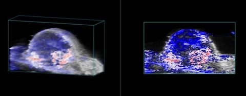
Oxygen saturation in tumor

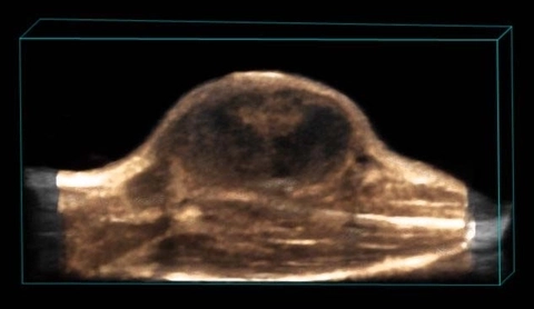
Perfusion in Tumor

3D rendered high-resolution ultrasound (greyscale) and nonlinear contrast (beige) image of a subcutaneous tumor showing perfusion in the tumor tissue.
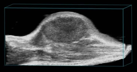
Subcutaneous tumor

3D rendered high-resolution ultrasound image of a subcutaneous tumor.

Nanoparticle distribution in tumor

High-resolution ultrasound (left) and spectrally unmixed photoacoustic (right) image of a subcutaneous tumor showing nanoparticle distribution (yellow) as well as oxygenated (red) and deoxygenated (blue) hemoglobin signal.

Perfusion in Tumor

High-resolution ultrasound (left) and nonlinear contrast (right) image of a subcutaneous tumor showing perfusion in the tumor tissue.
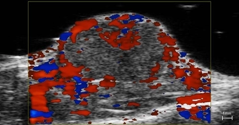
Blood flow in tumor

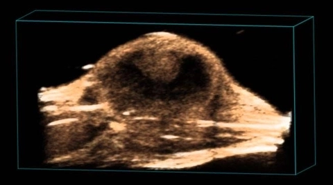
Perfusion in Tumor

3D rendered high-resolution nonlinear contrast image of a subcutaneous tumor showing perfusion in the tumor tissue.
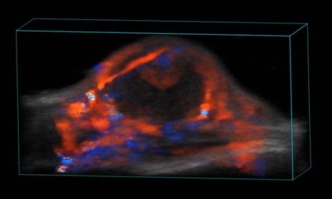
Blood flow in tumor

3D rendered high-resolution ultrasound (greyscale) and color Doppler (orange and blue) image of a subcutaneous tumor showing blood flow.
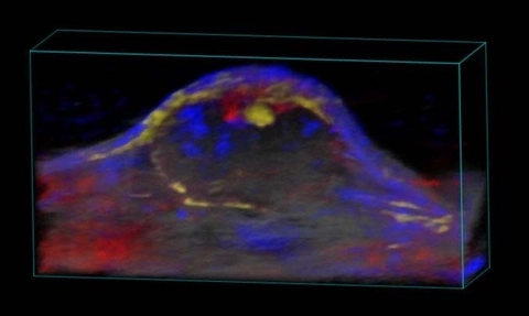
Nanoparticle distribution in tumor

3D rendered high-resolution ultrasound (greyscale) and spectrally unmixed photoacoustic (red, blue and gold) image of a subcutaneous tumor showing nanoparticle distribution (yellow) as well as oxygenated (red) and deoxygenated (blue) hemoglobin signal.


Mouse Subcutaneous Tumor with Contrast
Non-linear contrast bolus perfusion into a subcutaneous tumor in a mouse. See Oncology images.