Neurobiology
Neuroimaging Techniques for Seeing the Mouse Brain in vivo and in Real-time
Visualize functional processes for your small animal neuroscience research including stroke, cancer and in vivo delivery of drugs, stem cells, or other agents.
Compared to other preclinical neuroimaging techniques, Vevo imaging systems are non-invasive, non-ionizing, and offer a more cost effective solution.
Ultra-high frequency ultrasound and photoacoustic imaging is capable of assessing many neurologically relevant parameters in 2D and 3D such as:
- Blood flow
- Perfusion
- Oxygen saturation
- Total Hemoglobin
- Percent vascularity throughout the brain
- Contrast agents (ultrasound microbubbles, NIR dyes, nanoparticles)
In addition, the Vevo BRAIN stereotactic frame and neuroanatomical atlas facilitates mouse brain imaging for increased ease-of-use and reproducibility.
Elevate your neuroimaging research with VADA
Elevate your neuro-imaging research by utilizing the Vevo Advanced Data Acquisition (VADA) mode to access pre-beamformed, individual channel data. This open-architecture framework enables the implementation of custom plane wave transmit sequences, facilitating ultrafast acquisition and the collection of advanced vascular data. With VADA, you have the ability to assess cerebral microvasculature blood flow through ultrafast Doppler imaging or super resolution imaging.
Do you have a question?
Imaging the Brain
Vasculature in the Brain during Ischemic Stroke
Vasculature in the mouse brain, visualized with color Doppler during transient carotid artery occlusion.
Gallery

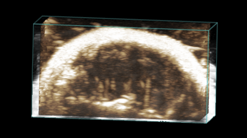
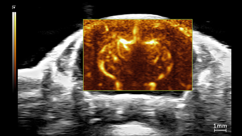
3D Non-linear Contrast of Mouse Brain
3D non-linear contrast view of a mouse brain. This was acquired using a UHF29x transducer on the Vevo F2xc imaging platform.
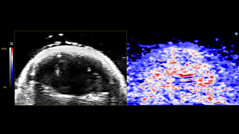
Oxy-Hemo View of Mouse Brain
Oxy-Hemo view of a mouse brain. This was acquired using a UHF29x transducer on the Vevo F2xc imaging platform.
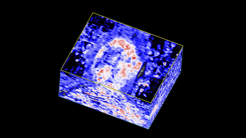
3D Oxy-Hemo View of Mouse Brain
3D Oxy-Hemo view of a mouse brain. This was acquired using a UHF29x transducer on the Vevo F2xc imaging platform.
Power Doppler view of Mouse Brain
Power Doppler view of a mouse brain. This was acquired using a UHF29x transducer on the Vevo F2xc imaging platform.
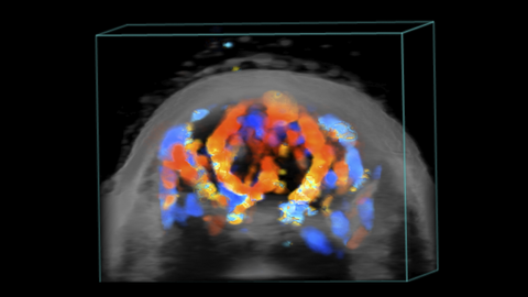
3D Power Doppler view of Mouse Brain
3D Power Doppler view of a mouse brain. This was acquired using a UHF29x transducer on the Vevo F2xc imaging platform.
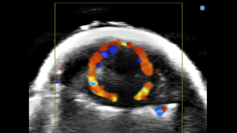
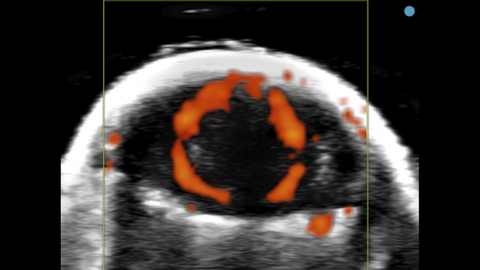
Color Doppler of Mouse Brain
Color Doppler view of a mouse brain. This was acquired using a UHF29x transducer on the Vevo F2xc imaging platform.
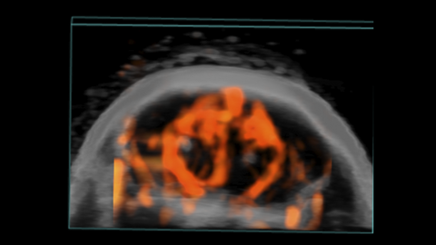
3D Color Doppler of Mouse Brain
3D Color Doppler view of a mouse brain. This was acquired using a UHF29x transducer on the Vevo F2xc imaging platform.
Ultrafast Doppler view of Mouse Brain
Ultrafast Doppler (UFD) view of the brain in a 1.5 month old CD-1 mouse. This was acquired using a UHF29x transducer on the Vevo F2xc imaging platform.
Publications
TOP PAPER
Ultrasound-Assisted Blood–Brain Barrier Opening Monitoring by Photoacoustic and Fluorescence Imaging Using Indocyanine Green
Ultrasound in Medicine and Biology
,
TOP PAPER
3D doppler ultrasound imaging of cerebral blood flow for assessment of neonatal hypoxic-ischemic brain injury in mice
PLoS ONE
,
TOP PAPER
In vivo imaging in experimental spinal cord injury – Techniques and trends
Brain and Spine
,
TOP PAPER
Early cerebrovascular and long-term neurological modifications ensue following juvenile mild traumatic brain injury in male mice
Neurobiology of Disease
,
TOP PAPER
Transcranial Photoacoustic Detection of Blood-Brain Barrier Disruption Following Focused Ultrasound-Mediated Nanoparticle Delivery
Molecular Imaging and Biology
,
TOP PAPER
Multimodal and multiscale optical imaging of nanomedicine delivery across the blood-brain barrier upon sonopermeation
Theranostics
,
TOP PAPER
Exploration of melanoma metastases in mice brains using endogenous contrast photoacoustic imaging
International Journal of Pharmaceutics
,
TOP PAPER
Very High Resolution Ultrasound Imaging for Real-Time Quantitative Visualization of Vascular Disruption after Spinal Cord Injury
Journal of Neurotrauma
, Stent treatment improves cerebral microcirculatory disorder and blood–brain barrier function in internal carotid artery stenosis via intercellular adhesion molecule 1 modulation
Journal of Cell Communication and Signaling
, Request a Quote or Demo