Biomarker and Molecular Imaging
Perform 2D or 3D Biomarker and Molecular Imaging in Small Animals
Biomarker research contributes significantly to breakthroughs in the understanding of disease progression as well as the validation of therapeutic treatments.
Using ultrasound and photoacoustic contrast agents with the Vevo systems, biomarkers can be non-invasively visualized deep within the anatomy of the animal in both 2D and 3D with resolutions down to 30 µm.
Published molecular imaging applications using the Vevo systems include biomarker quantification, monitoring drug delivery, cell tracking, diagnostics such as detection of metastatic cells, development and characterization of novel theranostic contrast agents.
In the drug development and theranostics space, the multi-modal nature of the Vevo systems make them ideal for assessing response to therapy including: morphology, tumor volume, angiogenesis, vascularity, perfusion and hypoxia as well as for assessing cardiotoxicity.
Molecular imaging can be performed on the Vevo imaging system using two categories of contrast agents:
Microbubbles (Ultrasound contrast)
- Non-toxic, micron-sized agents which stay within the vasculature
- Sensitivity down to the capillary level
- Contrast agents can be targeted or untargeted
- Truly translational - used in the clinic
Photoacoustic Contrast Agents
- Typically sub-100nm sized agents which can extravasate for tissue labeling
- Can be customized for a wide variety of targets and applications
- Can be multi-modal for PET, MR, Optical or other imaging compatability
Nonlinear Contrast Mode
Mouse Kidney Perfusion
Mouse Kidney Perfusion with Non-Targerted Microbubbles Imaged in Nonlinear Contrast Mode.
Nonlinear Contrast Mode
Tumor Perfusion - Displayed as MIP
Tumor Perfusion with Non-Targeted Microbubbles, Imaged with Nonlinear Contrast and Displayed as MIP.
Gallery
Nanoparticle distribution in tumor
3D rendered high-resolution ultrasound (greyscale) and spectrally unmixed photoacoustic (red, blue and gold) image of a subcutaneous tumor showing nanoparticle distribution (yellow) as well as oxygenated (red) and deoxygenated (blue) hemoglobin signal.
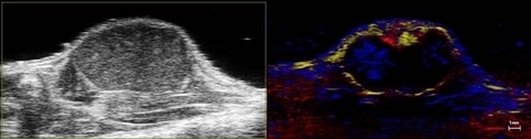
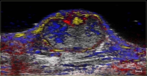
Nanoparticle distribution in tumor
High-resolution ultrasound (left) and spectrally unmixed photoacoustic (right) image of a subcutaneous tumor showing nanoparticle distribution (yellow) as well as oxygenated (red) and deoxygenated (blue) hemoglobin signal.
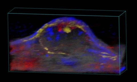
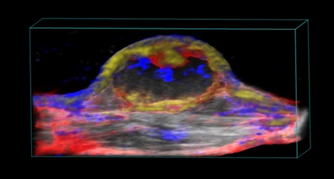
Nanoparticle distribution in tumor
3D rendered high-resolution ultrasound (greyscale) and spectrally unmixed photoacoustic (red, blue and gold) image of a subcutaneous tumor showing nanoparticle distribution (yellow) as well as oxygenated (red) and deoxygenated (blue) hemoglobin signal.

Stem cells in the mouse hindlimb
3D rendered spectrally unmixed photoacoustic image of a stem-cell injected, ischemic (right) and control (left) mouse hindlimb. Oxyhemoglobin (red), deoxyhemoglobin (blue) and dye-labelled stem cells (green) are shown.
Nanoparticle distribution in tumor
High-resolution ultrasound (greyscale) and spectrally unmixed photoacoustic (color) image of a subcutaneous tumor showing nanoparticle distribution (yellow) as well as oxygenated (red) and deoxygenated (blue) hemoglobin signal.
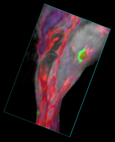
Mouse hindlimb showing blood and ICG
3D rendered high-resolution ultrasound (greyscale) and spectrally unmixed photoacoustic (red, blue and green) image of the mouse hindlbimb. The green color indicates ICG dye in the popliteal lymph node after a hindpaw injection, the red color indicated oxygenated hemoglobin and the blue color indicates deoxygenated hemoglobin.

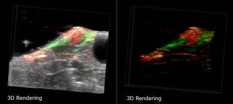
Breast Cancer Tumor in 3D
Breast Cancer Tumor in a mouse, imaged with photoacoustics in 3D. Shows localization of a targeted nanoparticle (green) within the tumor. Left panel shows both photoacoustics and ultrasound images, overlaid. Right panel shows only the photoacoustics signal.
Resources

Request a Quote or Demo