Vascular Biology
Preclinical Ultrasound for Vascular Biology Research
Visualize and Quantify Vascular Function
Ultra-high Frequency (UHF) Vevo imaging systems allow for non-invasive, real-time evaluation of vascular function with resolution down to 30 microns.
With UHF imaging and software, you can:
- Quantify pulse wave velocity and vascular resistance
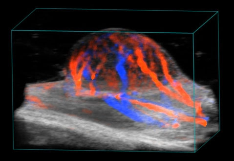
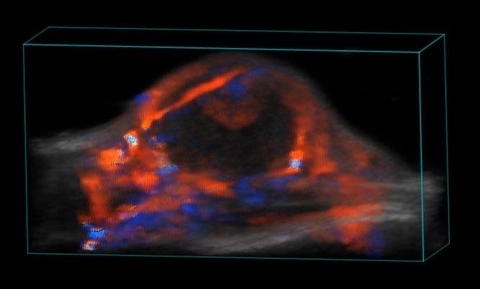
- Detect aneurysm and plaque formation with 3D reconstruction
- Analyze vessel wall characteristics (i.e. diameter, strain, velocity, stiffness, etc.)
- Quantify blood flow and perfusion
Perfect for Animal Models of:
- Atherosclerosis
- Aneurysm
- Arteriosclerosis
- Vascular Ischemia
Vascular Biology
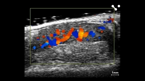
Right Femoral Artery Bifurcation in a Mouse
Gallery
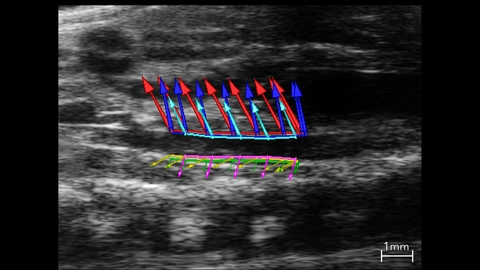
Abdominal Aorta Used In Strain Analysis
B-mode image of the abdominal aorta with vessel wall velocity vector arrows used in strain analysis.
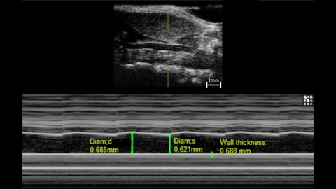
M-Mode of a Mouse Carotid Artery
M-mode image of mouse left common carotid.
Measurements: wall thickness and vessel diameter in systole and diastole.
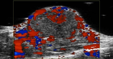
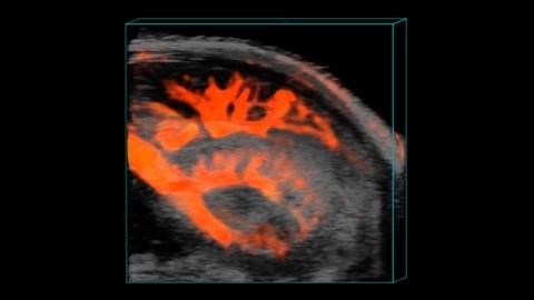
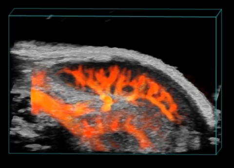
Splenic Vasculature in a Mouse

Publications
Request a Quote or Demo