LV Analysis
Trace measurements in B-Mode, M-Mode and EKV Mode
Trace measurements in B-Mode, M-Mode and EKV Mode using LV Analysis.
Clinically sourced functional and anatomical cardiovascular measurements in an easy-to-use tool.
The manual LV Analysis tool is widely adopted, having been available for well over 10 years across several different generations of Vevo Imaging Systems. Tracing measurements are available in B-Mode, M-Mode, and EKV Mode. The B-Mode / EKV Mode implementation adapts the clinically accepted modified Simpson’s monoplane method of disks approach for left ventricular analysis to small laboratory animals. These methodologies are discussed in the American Society Of Echocardiography consensus paper on Recommendations For Chamber Quantification1. In M-Mode, wall thicknesses and chamber dimensions across the sample line are used to calculate anatomical and functional parameters.
1. Lang RM, Bierig M, Devereux RB, et al. Recommendations for chamber quantification: A report from the American Society of Echocardiography’s guidelines and standards committee and the Chamber Quantification Writing Group, developed in conjunction with the European Association of Echocardiograph. J Am Soc Echocardiogr. 2005;18(12):1440-1463. doi:10.1016/j.echo.2005.10.005.
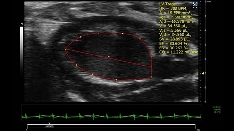
LV Trace of Parasternal Long Axis B-Mode image of mouse heart in full diastole
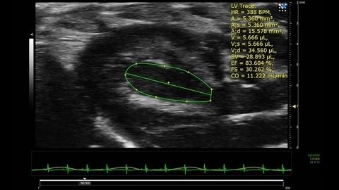
LV Trace of Parasternal Long Axis B-Mode image of mouse heart in full systole
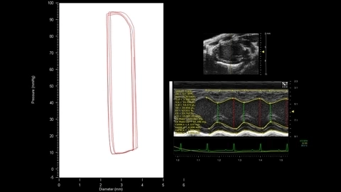
Pressure-Volume Loops
The Vevo Imaging Systems and animal handling equipment can be integrated with a third-party blood pressure monitoring system. Once connected and set up, blood pressure traces can then be displayed, and saved, as part of the physiological monitoring data. In conjunction with the LV Analysis tool, pressure-volume curves may be generated to determine pressure–volume relationships.
Discover Auto LV
Functional and anatomical analysis of the left ventricle in B-mode & M-mode with just one click!
Request a Quote or Software Trial