Dermatology Gallery
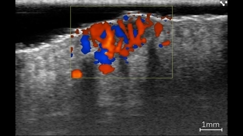
Color Doppler in Basal Cell Carcinoma Lesion
Color Doppler showing blood flow within a basal cell carcinoma lesion in a patient using ultra high frequency ultrasound on the Vevo MD.
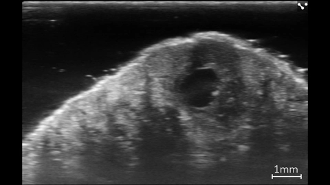
Basal Cell Carcinoma Lesion
High resolution image of a basal cell carcinoma lesion in a patient, imaged with the Vevo MD.
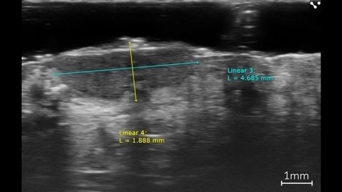
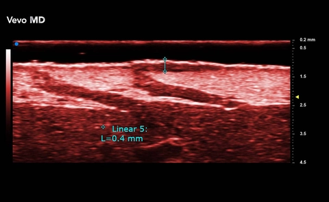
Linear Measurements of a Basal Cell Carcinoma
Linear measurements of a basal cell carcinoma in a patient using ultra high frequency ultrasound on the Vevo MD.
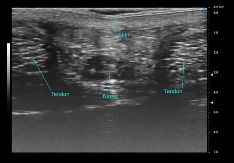
Palm - Adult Female
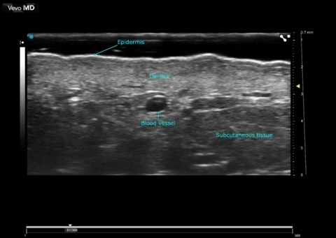
High Resolution Ultrasound Dermal Imaging
Scanned using a UHF70 transducer on the Vevo MD.
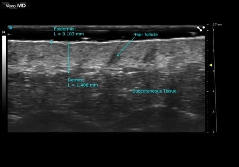
Facial Skin
Ephelis
Ephelis

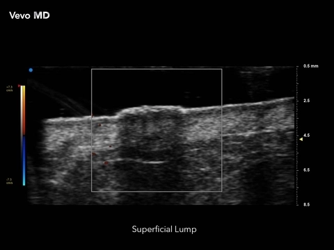
Superficial Lump
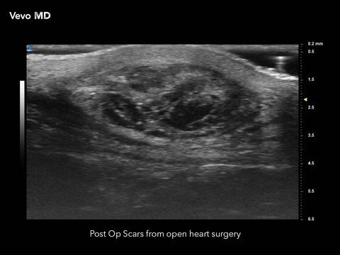
Post Op Scars
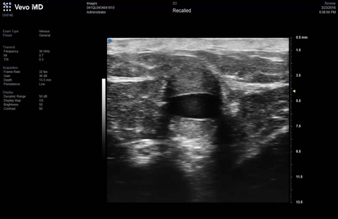
Vein with Valve
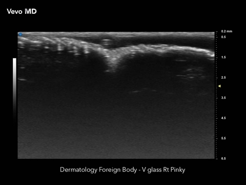
Foreign Body
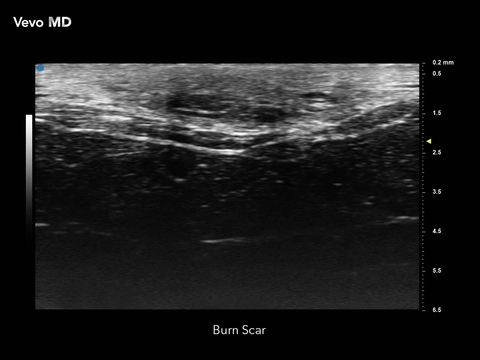
Burn Scar