Fibrosis and Tissue Stiffness Gallery
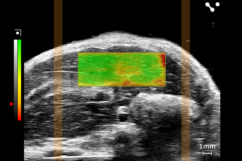
Shear Wave Elastography: Wide Box in Healthy Liver
Showing the wide SWE box used in a healthy mouse liver on the UHF29x.

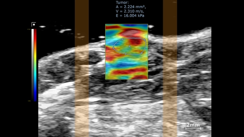
Stiffness map in tumor region
SWE mode overlay showing stiffness map in the tumor region. This was acquired using a UHF57x transducer on the Vevo F2xc imaging platform.
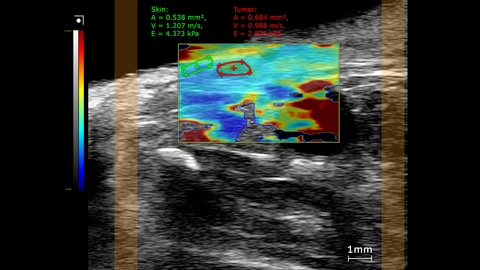
Subcutaneous prostate tumor stiffness map
SWE mode stiffness map showing elastography measurements comparing the tumor to skin inside a mouse. This image was obtained using a UHF29x transducer on a Vevo F2xc imaging platform.
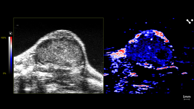
Oyx-hemo Imaging of Mouse Tumor
Oxy-hemo image of a mouse tumor showing shear wave elastography, scanned using a UHF29x transducer.

Contrast Imaging of Mouse Tumor
Contrast image of a mouse tumor showing shear wave elastography, scanned using a UHF29x transducer.
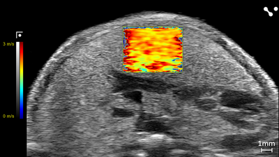
Shear Wave Elastography: NAFLD/MASLD Mouse - Velocity
Liver in a NAFLD/MASLD Mouse showing Velocity (m/s) through Shear Wave Elastography scanned using a UHF29x transducers on a Vevo F2 system.
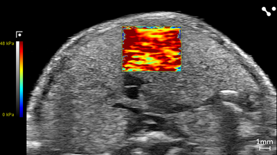
Shear Wave Elastography: NAFLD/MASLD Mouse - Stiffness
Liver in a NAFLD/MASLD Mouse showing Stiffness (kPa) through Shear Wave Elastography scanned using a UHF29x transducers on a Vevo F2 system.
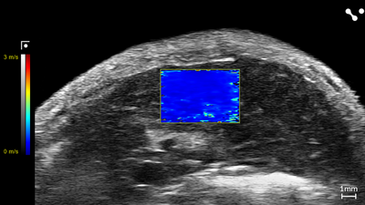
Shear Wave Elastography: Control Mouse - Velocity
Liver in a Control Mouse showing Velocity (m/s) through Shear Wave Elastography scanned using a UHF29x transducers on a Vevo F2 system.
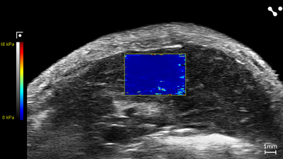
Shear Wave Elastography: Control Mouse - Stiffness
Liver in a Control Mouse showing Stiffness (kPa) through Shear Wave Elastography scanned using a UHF29x transducers on a Vevo F2 system.