Cardiovascular Biology Gallery

Aortic Arch in a Mouse


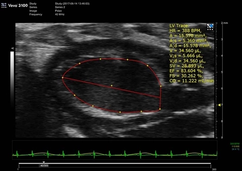
LV Trace of Parasternal Long Axis
B-Mode image of mouse heart in full systole.
PSLAX LV Trace B-Mode Diastole
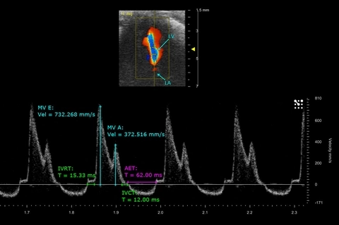
Mitral flow and measurements of a mouse neonate heart
PW Doppler of mitral blood flow in a postnatal day 9 mouse taken from an apical 4 chamber view, with related cardio measurements.
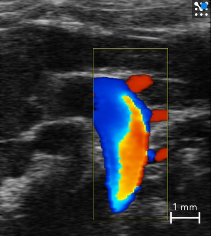
Mouse Aortic Arch
Color Doppler image of the aortic arch in a mouse showing all three branches.
Oxygen saturation in the myocardium
High-resolution ultrasound (greyscale) and photoacoustic (red, white and blue) image of a long axis view of the left ventricle of the mouse heart. The photoacoustic image is a parametric map of oxygen saturation in the myocardium.
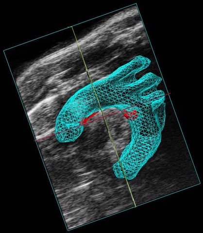
Atherosclerosis in the Aortic Arch
3D volume reconstruction of the mouse aortic arch (in blue) with atherosclerotic plaques (in red).
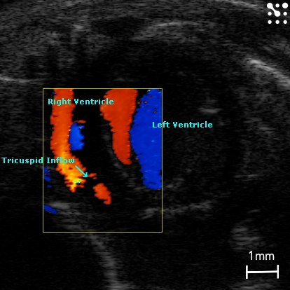
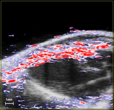
Tricuspid Flow in the Mouse
Apical four chamber view illustrating blood flow through the tricuspid valve, imaged with color Doppler.
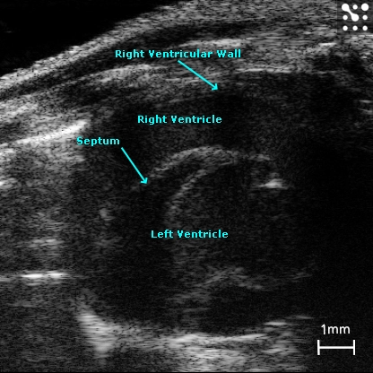
Mouse Right Ventricle
The right ventricle in a mouse imaged in B-Mode.