Dermatology
High-Frequency Ultrasound for Imaging Superficial Anatomy
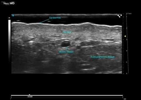
Imaging the skin layer is often difficult with conventional ultrasound.
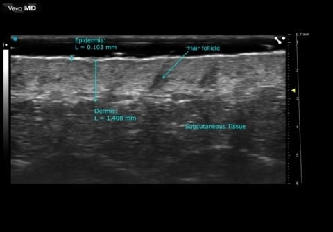
The Vevo MD is the world’s first dermatology ultrasound system designed specifically for imaging superficial anatomy; ideally suited to image the following dermatological applications:
- Skin layers
- Melanoma
- Lipomas
- Hair follicles (hair loss)
- Foreign Body Identification
- Lumps and Bumps
Gallery
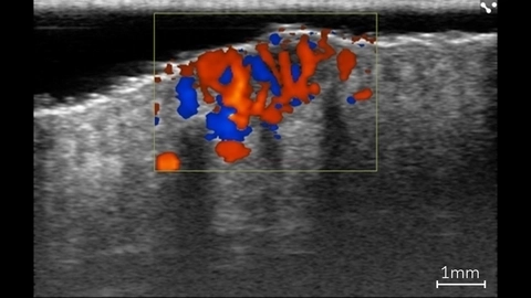
Color Doppler in Basal Cell Carcinoma Lesion
Color Doppler showing blood flow within a basal cell carcinoma lesion in a patient using ultra high frequency ultrasound on the Vevo MD.
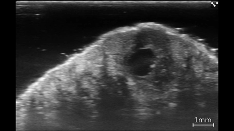
Basal Cell Carcinoma Lesion
High resolution image of a basal cell carcinoma lesion in a patient, imaged with the Vevo MD.
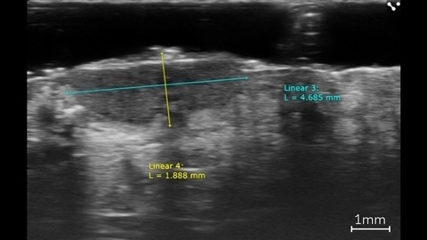
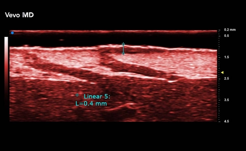
Linear Measurements of a Basal Cell Carcinoma
Linear measurements of a basal cell carcinoma in a patient using ultra high frequency ultrasound on the Vevo MD.
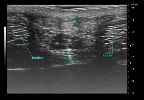
Palm - Adult Female
High Resolution Ultrasound Dermal Imaging
Scanned using a UHF70 transducer on the Vevo MD.
Facial Skin
Ephelis
Ephelis

Publications
TOP PAPER
Overview of Ultrasound Imaging Applications in Dermatology
Journal of Cutaneous Medicine and Surgery
,
TOP PAPER
Ultra-High Frequency Ultrasound, A Promising Diagnostic Technique: Review of the Literature and Single-Center Experience
Canadian Association of Radiologists Journal
,
TOP PAPER
A Preliminary Study for Quantitative Assessment with HFUS (High- Frequency Ultrasound) of Nodular Skin Melanoma Breslow Thickness in Adults Before Surgery: Interdisciplinary Team Experience
Current Radiopharmaceuticals
,
TOP PAPER
Seventy‐MHz Ultrasound Detection of Early Signs Linked to the Severity, Patterns of Keratin Fragmentation, and Mechanisms of Generation of Collections and Tunnels in Hidradenitis Suppurativa
Journal of Ultrasound in Medicine
,
TOP PAPER
Performance of ultra-high-frequency ultrasound in the evaluation of skin involvement in systemic sclerosis: a preliminary report
Rheumatology
, Seeing More to Treat Better: Ultra-High Frequency Ultrasound as a Decision-Shaping Tool in Radiotherapy for Head and Neck Non-Melanoma Skin Cancer in a Single-Institution Feasibility Study
Cancers
, Ultra-High-Frequency Ultrasound of Melanoma Excision Scars for Detection of Clinically Occult Local Recurrence: A Single-Center Retrospective Study
Clinical, Cosmetic and Investigational Dermatology
, High- and Ultra-High-Frequency Ultrasound Identifies a Subclinical Link Between Suppurative Comedonal Nevus and Hidradenitis Suppurativa
Journal of Ultrasound in Medicine
, Ultrasound Patterns of Vitiligo at High Frequency and Ultra-High Frequency
Journal of Ultrasound in Medicine
, Request a Quote or Demo