This recent article by Lavaud et al. uses high-frequency ultrasound and spectroscopic photoacoustic imaging (PAI), along with commercially available contrast agents to non-invasively examine metastatic liver lesions.
A conversation with Dr Florian Raes. He shares the development of an all-in-one multimodal imaging platform combining the Vevo LAZR’s ultrasound and photoacoustic capabilities with bioluminescence and near-infrared fluorescence.
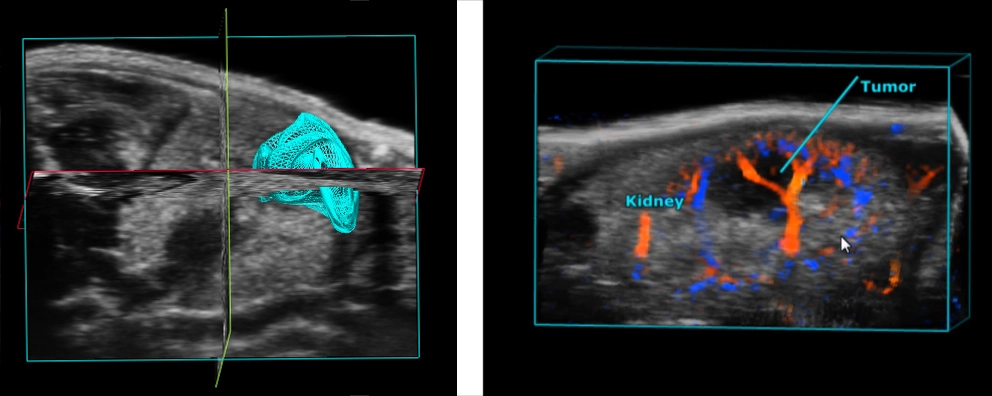
In this study, Ramasawmy et al.1 sought to compare 9 Tesla (T) MRI, Benchtop (1T) MRI, Vevo US and BLI in the ability to monitor and detect tumor growth in a xenograft colorectal metastasis model in the mouse liver.
In this talk, recorded during the Innovation in Imaging Summit 2018 in Toronto, researcher Danielle Charron shares her lab's approach to creating photoacoustic nanoparticles. She highlights how a lot of the work they do in fluorescent imaging can also be done using photoacoustics as well.
This study by Greco et al. highlights the benefits of using Ultra High Frequency Ultrasound-guided injection versus surgery to generate an orthotopic mouse model of thyroid cancer.
Watch this very interesting presentation (by Dr. Florian Raes) that shares research aimed at assessing tumor oxygenation to validate non-invasive imaging measurements of tumor hypoxia.
This recent article titled by Lavaud et al., uses high-frequency ultrasound and spectroscopic photoacoustic imaging as a multimodal non-invasive examination tool for the assessment of melanoma brain tumors in an orthotropic mouse model.
Metastasis rather than primary tumors determine prognosis and mortality in the majority of cancer patients. Detection of metastatic lesions is critical to determine prognosis and suitable treatments. We developed a method... Read full story.
This recent article by Bruckner et al., uses commercially available target-ready microbubbles and antibodies to non-invasively assess the severity of intestinal inflammation and development of tumors in a mouse model of colitis.
This featured publication by Shirinfard et al, uses a novel microbubbles injection technique to evaluate the feasibility of using dynamic contrast enhanced ultrasound (CEUS) to assess absolute perfusion of tissue in mice.
CRISPR-Cas 9 is a method used to target and edit DNA at precise locations in living cells. This has broad implications in treating human disease, specifically in the field of oncology. Read full blog.
This article by Wilson, et al. uses PA contrast agent with an antibody specific to breast cancer cells to perform in vivo molecular imaging in an orthotopic mouse model of breast cancer.
Molecular Imaging is emerging as an innovative and multidisciplinary field of research aimed at visualizing and quantifying the signature of diseases, making it possible for an earlier and more precise diagnosis. Read more.