Vevo F2xc Gallery

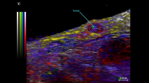
Unmixing from a Spectro scan of a subcutaneous prostate tumor
Unmixing from a Spectro scan of the Subcutaneous prostate tumor and surrounding region. This was acquired using a UHF57x transducer on the Vevo F2xc imaging platform.
Colormap – red/blue = oxy/deoxyhemo; yellow = lipid, green = collagen (theoretical).
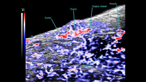
OxyHemo sO2 map of a subcutaneous prostate tumor
PA mode scans showing an OxyHemo sO2 map of a subcutaneous prostate tumor. This image was acquired using a UHF57x transducer on the Vevo F2xc imaging platform.
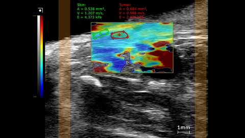
Subcutaneous prostate tumor stiffness map
SWE mode stiffness map showing elastography measurements comparing the tumor to skin inside a mouse. This image was obtained using a UHF29x transducer on a Vevo F2xc imaging platform.
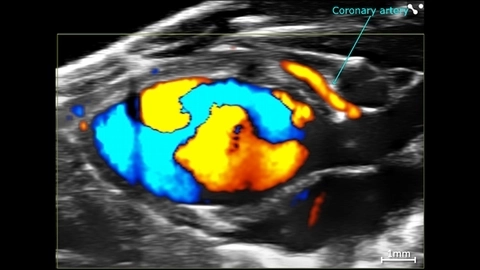
Cardiac Flow Dynamics and Coronary Artery
Cardiac flow dynamics and Coronary Artery visualized with Color Doppler EKV on the Vevo F2 System.

Canine eye in vivo
Entire canine eye visualized in vivo with the Vevo F2, scanned using a UHF22x transducer.
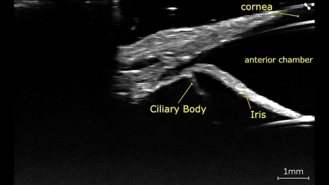
Canine eye angle of the anterior chamber
Canine eye imaged in vivo on the Vevo F2 scanned using the UHF71x.
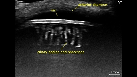
Canine eye, Ciliary body and processes
In vivo canine eye with ciliary processes, imaged with high-frequency on the Vevo F2 scanned using the UHF71x.
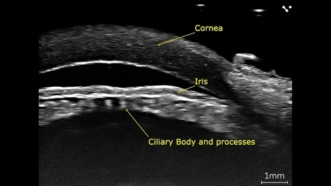
Porcine eye, Ciliary body and processes
Porcine eye imaged ex-vivo using the Vevo F2 with a UHF71x transducer.
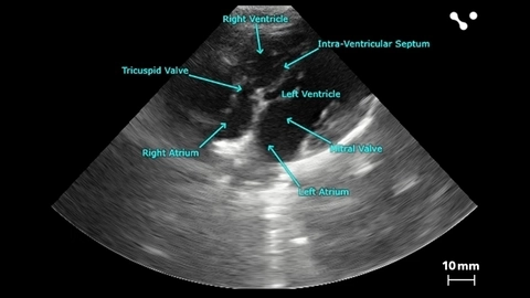
Apical 4 Chamber View of a Beagle
Apical 4 chamber view of a beagle scanned using a P5-1 transducers on the Vevo F2. Images courtesy of Drs. Kenneth Hoyt and Jay Griffin at Texas A&M University.
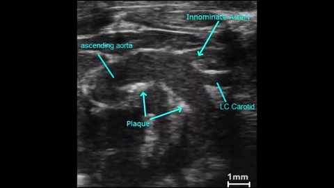
Atherosclerotic Plaque in an Aortic Arch
2D image showing atherosclerotic plaques in a mouse aortic arch.
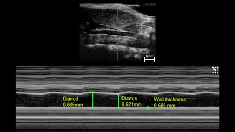
M-Mode of a Mouse Carotid Artery
M-mode image of mouse left common carotid.
Measurements: wall thickness and vessel diameter in systole and diastole.
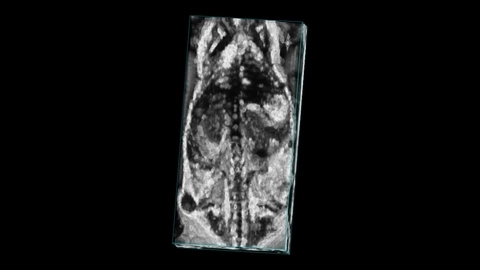
Mouse whole body with tumor
Whole body image of a mouse with a subcutaneous tumor visible on the flank, imaged with high frequency ultrasound on the Vevo F2 system.
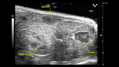
Ovary, spleen and kidney
B-mode image of the ovary, spleen and kidney, acquired using a UHF57x transducer on the Vevo F2.
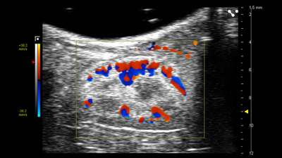
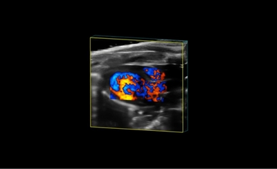
Sagittal View in the Left Kidney
Color Doppler image of the sagittal view in the left kidney, acquired using a UHF57x transducer on the Vevo F2.
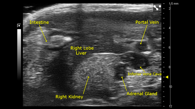
Right Liver Lobe and Adrenal Gland
B-mode image of the right liver lobe and adrenal gland, acquired using a UHF57x transducer on the Vevo F2.