Urogenital Gallery

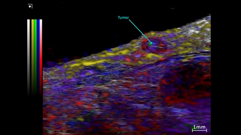
Unmixing from a Spectro scan of a subcutaneous prostate tumor
Unmixing from a Spectro scan of the Subcutaneous prostate tumor and surrounding region. This was acquired using a UHF57x transducer on the Vevo F2xc imaging platform.
Colormap – red/blue = oxy/deoxyhemo; yellow = lipid, green = collagen (theoretical).
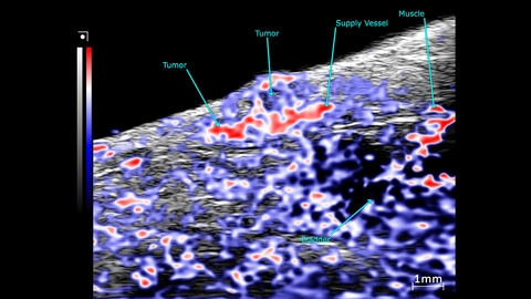
OxyHemo sO2 map of a subcutaneous prostate tumor
PA mode scans showing an OxyHemo sO2 map of a subcutaneous prostate tumor. This image was acquired using a UHF57x transducer on the Vevo F2xc imaging platform.
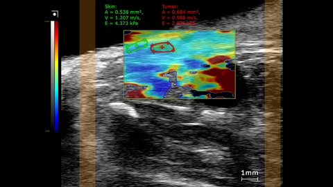
Subcutaneous prostate tumor stiffness map
SWE mode stiffness map showing elastography measurements comparing the tumor to skin inside a mouse. This image was obtained using a UHF29x transducer on a Vevo F2xc imaging platform.
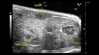
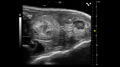
Ovary, spleen and kidney
B-mode image of the ovary, spleen and kidney, acquired using a UHF57x transducer on the Vevo F2.
Mouse ovary and kidney
B-mode image of the mouse ovary and kidney, acquired using a UHF57x transducer on the Vevo F2.
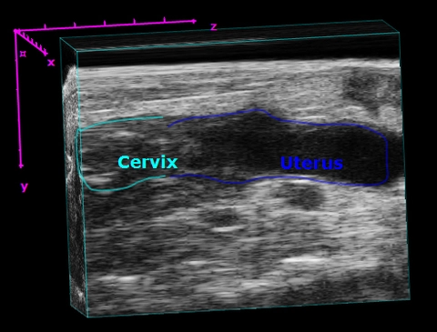
Mouse Cervix and Uterus
3D visualization of the mouse cervix and uterus from a sagittal view.
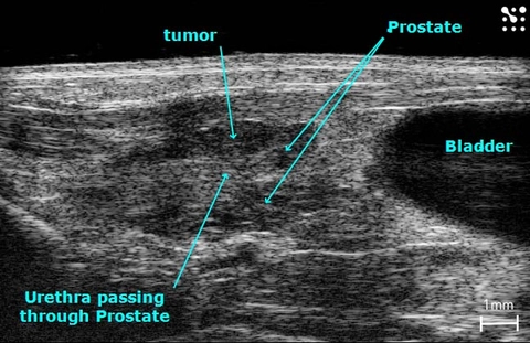
Murine Prostatic Tumor
B-Mode image of a prostate tumor in an adult mouse with the prostate, tumor and surrounding structures labelled.
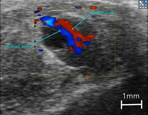
Ovarian Artery and Vein in a Mouse
Color Doppler image of the ovarian artery and vein in a mouse.