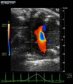
With the increased prevalence of cardiac diseases associated with preserved systolic function (i.e. HFPEF), diastolic functional measurements are becoming increasingly important. Read more.
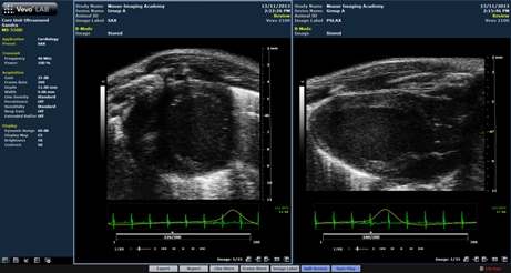
The split screen function, which is available on all current Vevo systems and in VevoLab is a great tool for longitudinal studies and for image optimization. Read more.
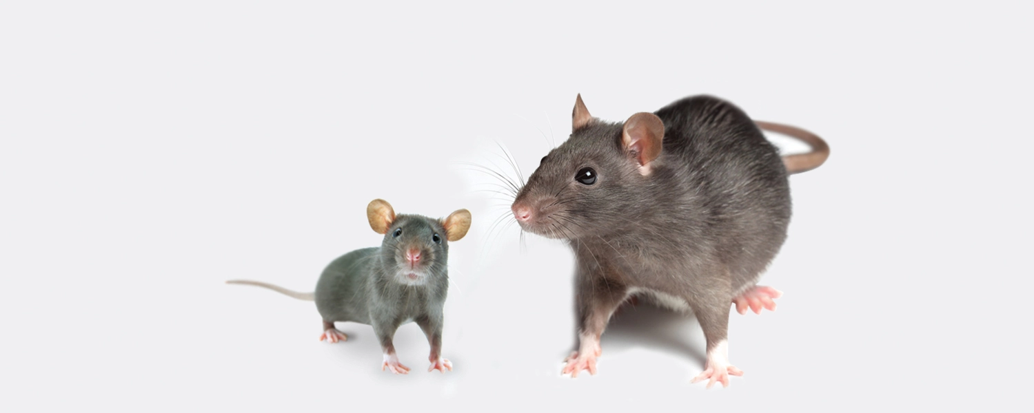
Although you would originally assume that echocardiography would be easier in rat than in mouse, anyone who has done both will quickly acknowledge that the former is actually more challenging. Find out more.
We invite you to view this previously recorded webinar presented by our guest speakers, Dr. Craig Goergen and Arvin Soepriatna from the Weldon School of Biomedical Engineering at Purdue University in West Lafayette, Indiana.
This webinar is presented by Andrew Needles, Senior Manager, Product Innovation, FUJIFILM, VisualSonics, Inc. This webinar explores the capabilities of the Vevo 3100 Ultrasound Imaging System for preclinical research across cardiovascular, cancer, developmental, abdominal and neurobiology
Take a look at the NEW Vevo LAZR-X Photoacoustic Imaging System (featured at WMIC - World Molecular Imaging Congress in September). Presented by Drew Heinmiller, Product Manager, Fujifilm VisualSonics.
This webinar is hosted by Mathew Platt, B.Sc from Dr. Jeremy Simpson's laboratory, University of Guelph. An improved non-invasive method for evaluating post mycoardial infarction cardiac output in a murine model.
Presented by Stephen Buttars, MSc, Product Manager at FUJIFILM VisualSonics. This webinar showcases how to measure cardiac wall strain using Vevo Strain. Stephen shares relevance of strain and strain rate measures, conventional functional measures vs. vevo strain analysis, recent publications and software updates.
Preclinical in vivo studies is a critical step in drug development. In vivo imaging in such studies can benefit drug development with rapid and predictive assays to assess drug toxicity and efficacy to reduce clinical attrition rates. High frequency ultrasound imaging will be addressed in this regard by the panel of speakers.
Learn: How to do a mouse echocardiogram, including imaging the heart in long axis, short axis and apical views, how to obtain images of the aortic valve, pulmonary valve, mitral and tricuspid valves, how to use Color Doppler Mode and much more. Click to learn more.