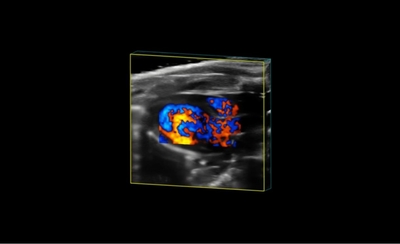
Image Gallery 4D-Mode Imaging Gallery Parasternal Long Axis in Color 4D Parasternal Long Axis in Color 4D Mouse Abdominal Aorta in 4D Mouse Abdominal Aorta in 4D The mouse abdominal aorta imaged in 4D from a longitudinal view. Cardiac Hypertrophy in 4D Cardiac Hypertrophy in 4D A model of murine cardiac hypertrophy imaged using 4D. Image courtesy of Dr. Craig Goergen, Purdue University. Left-Ventricular Myocardial Hypertrophy in 4D Left-Ventricular Myocardial Hypertrophy in 4D 4D image of mouse left ventricle (long and short axis views), displaying evident myocardial hypertrophy. 4D Image of the Mouse Left Ventricle 4D Image of the Mouse Left Ventricle 4D image of the mouse left ventricle, acquired from a parasternal short axis view. 4D Image of the Mouse Right Atrium 4D Image of the Mouse Right Atrium 4D image of the right atrium in a mouse. 4D Image of the Mouse Left Ventricle 4D Image of the Mouse Left Ventricle 4D image of the mouse left ventricle, acquired from a parasternal long axis view. 4D Image of the Mouse Left Atrium 4D Image of the Mouse Left Atrium 4D image of the left atrium in a mouse. 4D Left Ventricular Volume Overlay 4D Left Ventricular Volume Overlay 4D image of the mouse left ventricle acquired from a parasternal short axis view. A 4D volume was drawn and can be visualized in red.